 Skip to content
FOOT AND ANKLE / POSTOPERATIVE PROTOCOL
Calcaneal fracture — rehabilitation after fixation
Adults after fixation of a displaced calcaneal fracture, particularly an intra-articular fracture, without concurrent fusion. Skin condition, the subtalar joint surface and fixation stability are central.
For clinicians, physiotherapists and patients. The individual plan based on the operative report takes precedence over the time ranges on this page.
Essential protection
- Weight-bearing
- In a conventional protective pathway, weight-bearing is commonly deferred until around 8–12 weeks, depending on comminution, stability and union. Earlier progression requires a specific surgeon-prescribed plan.
- Brace / immobilisation
- Splint or boot; protect the wound and avoid heel pressure. An ordinary walking boot does not unload the heel like a specialist orthosis.
- Range of motion
- Gentle ankle and preserved subtalar movement may begin early if the wound and surgeon permit. Motion does not authorise loading.
Rehabilitation at a glance
Timings are approximate. Progression requires criteria and clearance for the relevant motion or loading level.
- 01
Skin care and protection
Weeks 0–2Protect heel skin, control swelling and enable safe transfers.
- 02
Movement while unloaded
Weeks 2–8/12Limit avoidable stiffness while protecting the bone.
- 03
Gradual loading
After clearance, often from weeks 8–12Restore walking without overloading the heel.
- 04
Strength and everyday function
Usually from weeks 12–16; later with complicationsImprove tolerance of standing, walking and uneven ground.
Full rehabilitation board
Click to enlarge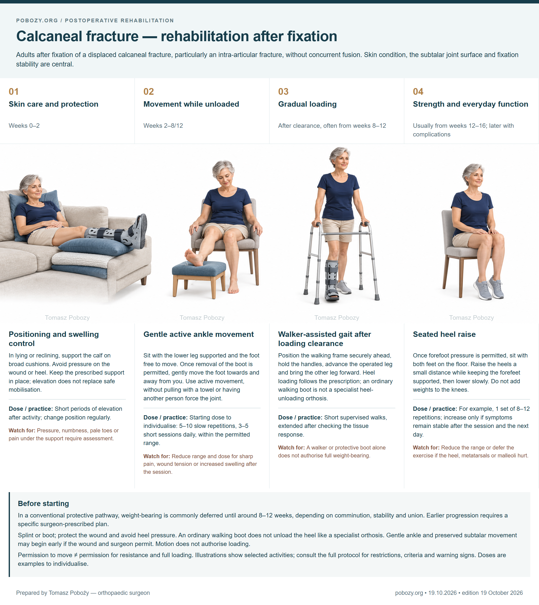
Exercises — take a closer look4 phases • technique • dosage • when to stop
Each phase has its own illustration. The descriptions below are the text equivalent of the board. Doses are starting examples to individualise with a physiotherapist and do not override surgical restrictions.
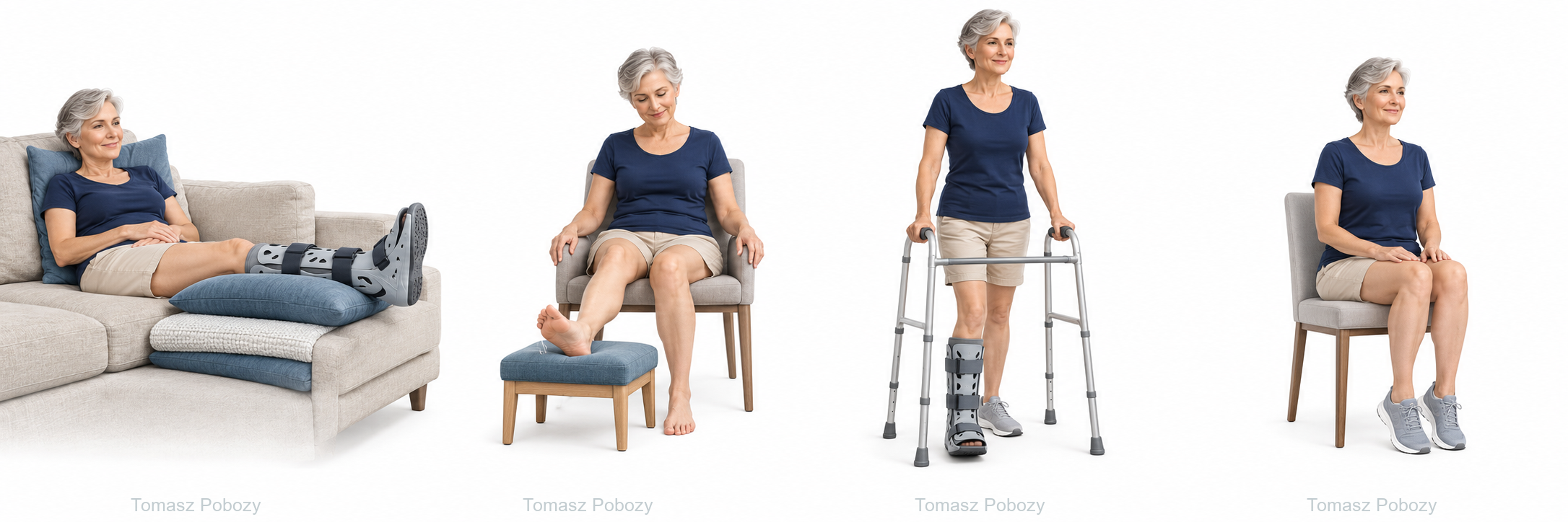
Positioning and swelling control
In lying or reclining, support the calf on broad cushions. Avoid pressure on the wound or heel. Keep the prescribed support in place; elevation does not replace safe mobilisation.
- Dose to individualise
- Short periods of elevation after activity; change position regularly.
- When to reduce or stop
- Pressure, numbness, pale toes or pain under the support require assessment.
Gentle active ankle movement
Sit with the lower leg supported and the foot free to move. Once removal of the boot is permitted, gently move the foot towards and away from you. Use active movement, without pulling with a towel or having another person force the joint.
- Dose to individualise
- Starting dose to individualise: 5–10 slow repetitions, 3–5 short sessions daily, within the permitted range.
- When to reduce or stop
- Reduce range and dose for sharp pain, wound tension or increased swelling after the session.
Walker-assisted gait after loading clearance
Position the walking frame securely ahead, hold the handles, advance the operated leg and bring the other leg forward. Heel loading follows the prescription; an ordinary walking boot is not a specialist heel-unloading orthosis.
- Dose to individualise
- Short supervised walks, extended after checking the tissue response.
- When to reduce or stop
- A walker or protective boot alone does not authorise full weight-bearing.
Seated heel raise
Once forefoot pressure is permitted, sit with both feet on the floor. Raise the heels a small distance while keeping the forefeet supported, then lower slowly. Do not add weights to the knees.
- Dose to individualise
- For example, 1 set of 8–12 repetitions; increase only if symptoms remain stable after the session and the next day.
- When to reduce or stop
- Reduce the range or defer the exercise if the heel, metatarsals or malleoli hurt.
Who this protocol is for
Adults after fixation of a displaced calcaneal fracture, particularly an intra-articular fracture, without concurrent fusion. Skin condition, the subtalar joint surface and fixation stability are central.
Subtalar fusion, tuberosity avulsion involving the Achilles insertion, threatened skin, infection and open injury require a separate plan. Do not mobilise a fused joint.
Before starting, confirm the operation date and extent, fixation, permitted loading, support-removal rules, range of motion and review date. Obtain any missing instruction from the surgeon before progressing.
Full programme by phase
01 · Skin care and protectionWeeks 0–2
- Goal
- Protect heel skin, control swelling and enable safe transfers.
- Weight-bearing
- Non-weight-bearing in the baseline protective pathway. Select crutches, a walker or wheelchair according to ability.
- Support / immobilisation
- Protective splint; check pressure areas. Avoid sustained support directly beneath the heel or wound.
- Motion
- Move the knee and hip freely. Ankle/subtalar motion may be allowed in the first few days, but only if tissue condition permits and the surgeon prescribes it.
- Work in this phase
- Calf elevation, position changes, thigh exercises and fall prevention.
- Progression criteria
- Wound assessed without threatened skin, swelling controlled and clear movement and protection instructions.
02 · Movement while unloadedWeeks 2–8/12
- Goal
- Limit avoidable stiffness while protecting the bone.
- Weight-bearing
- Remain non-weight-bearing until scheduled review and clearance. Do not shorten protection independently, even if movement is painless.
- Support / immobilisation
- Boot or other support; removal for exercise and hygiene as prescribed.
- Motion
- Gentle active ankle motion; small subtalar movements once cleared. Avoid forceful manipulation, Achilles stretching and resistance through the heel.
- Work in this phase
- Short movement sessions, hip and knee training and independent mobility without heel loading.
- Progression criteria
- Assessment of union imaging, wound, pain and stability supports a decision on when and how much to load.
03 · Gradual loadingAfter clearance, often from weeks 8–12
- Goal
- Restore walking without overloading the heel.
- Weight-bearing
- Increase loading in stages under a written plan. Practise any kilogram limit with a physiotherapist; do not set loading increments independently.
- Support / immobilisation
- Boot and walking aids, followed by cushioned footwear once cleared.
- Motion
- Gradually increase active range and control; do not force painful inversion or eversion.
- Work in this phase
- Walker- or crutch-assisted gait and swelling checks after activity. Heel raises require permission for forefoot loading and calf work.
- Progression criteria
- Walking at the permitted load without next-day worsening, continued union and clearance for strengthening.
04 · Strength and everyday functionUsually from weeks 12–16; later with complications
- Goal
- Improve tolerance of standing, walking and uneven ground.
- Weight-bearing
- Begin with gentle tasks; return to standing work and sport may take many months. Running requires a separate assessment.
- Support / immobilisation
- Well-fitting shoes; orthotics when indicated. Check for rubbing if heel shape has changed.
- Motion
- Functional range without forcing the subtalar joint. Persistent pain or stiffness may require assessment of post-traumatic changes.
- Work in this phase
- Seated heel raises, progressing to standing when cleared, balance and graded outdoor tasks.
- Progression criteria
- Union, acceptable task tolerance, no marked next-day flare and safe limb control.
How to adjust the dose
Increase one element at a time: repetitions, distance, range or resistance. Sharp pain, limping or markedly increased swelling calls for reducing the load. Check symptoms after exercise and the following day; discuss persistent deterioration. Absence of pain does not replace union assessment.
If the surgeon prescribes a kilogram limit, record the value and duration. Do not independently convert “as tolerated” into kilograms. Do not add weights without clearance for resistance.
Everyday activities and return to function
- Walking and stairs
- Use aids while needed to respect the load restriction and maintain gait quality. Practise stairs with a physiotherapist; choose a safer alternative when support or a rail is unavailable.
- Driving
- Resume only after assessment of rapid, forceful emergency braking and without medication impairing alertness. Operated side, transmission, bracing and loading restrictions affect the decision; discuss it with the treating team and consider insurance conditions.
- Work, rest and swelling
- Seated work requires opportunities to change position and elevate the leg; standing or manual work requires loading and task tolerance. Swelling may persist, but new or rapidly increasing swelling requires assessment.
- Wound care and prevention
- Do not soak an unhealed wound, remove wires yourself or change prescribed thromboprophylaxis. Follow individual medication, dressing, compression and scar-care instructions. Swimming requires complete wound closure and clearance.
- Running and sport
- Timing follows healing and capacity; it is not a stand-alone criterion. Establish safe gait, strength, balance and task tolerance first, progressing without significant next-day worsening.
Warning signs — when to seek help
- Emergency help
- Sudden shortness of breath, chest pain, collapse or coughing up blood: call emergency services. Use 112 in the EU; elsewhere use the appropriate local emergency number.
- Urgent same-day assessment
- New one-sided calf pain and swelling; increasing pain despite rest; fever with wound redness or discharge; wound breakdown; a new injury or pain after accidental loading.
- Do not wait for a routine appointment
- A cold, pale or blue foot, rapidly increasing numbness, loss of toe movement or severe increasing pain under a cast requires immediate emergency assessment.
Variants and clinical context
- Early motion and early weight-bearing are separate decisions. Skin condition may delay even gentle exercise.
- The 2026 study used a specialist heel-unloading orthosis after an initial 4 non-weight-bearing weeks and was retrospective. It does not justify immediate full weight-bearing or use of an ordinary boot for that purpose.
- If subtalar fusion was performed, use the fusion pathway; do not exercise to restore motion at the fused joint.
Related resources
References and evidenceVerified publications and evidence limitations
- Operative versus non-operative treatment for closed, displaced, intra-articular fractures of the calcaneus: randomised controlled trial. The BMJ. 2014. DOI: 10.1136/bmj.g4483.
- Early Full Weight-Bearing After Calcaneal Fracture Using a Heel-Unloader Orthosis: Equivalent Radiographic Outcomes Compared to Traditional Cast Treatment. Foot & Ankle Orthopaedics. 2026. DOI: 10.1177/24730114261434237.
Research informs protection and progression decisions; it does not validate every illustrated exercise or example dose. Procedure details, tissue condition and the individual plan determine application.
Glossary
- ROM
- Joint range of motion. Active movement uses your own muscles; assisted movement uses additional help.
- Non-weight-bearing
- No body-weight transfer through the operated foot during standing and walking.
- As tolerated
- Once this pathway is authorised: loading limited by symptoms and movement quality within prescribed protection. It is not permission for unrestricted sport.
- Walker
- A walking boot is a protective ankle-foot support. A walking frame is a separate mobility aid.