 Skip to content
FOR DOCTORS AND PHYSIOTHERAPISTS
Musculoskeletal Ultrasound
Professional resources on musculoskeletal ultrasound: protocols, anatomy, pathology, dynamic examination and diagnostic pitfalls.
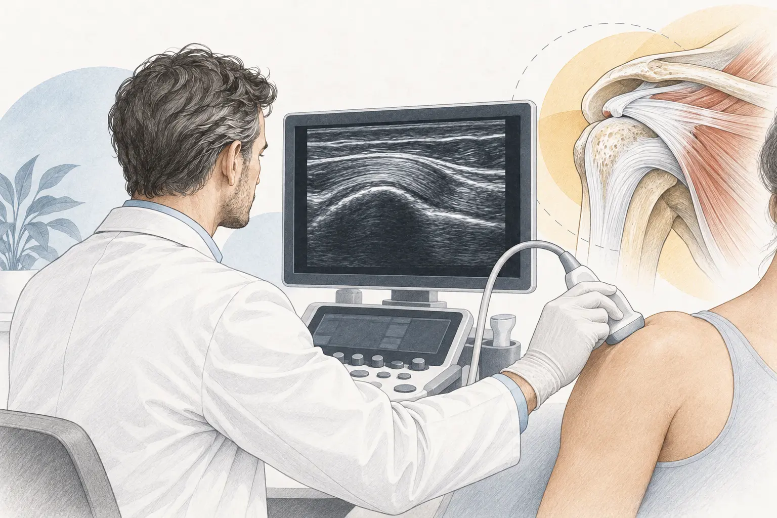
Musculoskeletal ultrasound should answer a specific clinical question. Moving the probe over successive structures is less useful than a hypothesis-driven examination that continuously relates the ultrasound findings to the history, symptom location and functional assessment.
How an orthopaedic surgeon structures an ultrasound examination
1. Clinical question
Before starting, identify the most likely diagnoses and which structure may explain the patient’s symptoms.
2. Examination using anatomical landmarks
Consistent anatomical orientation and a reproducible imaging sequence help reduce missed structures and make serial examinations easier to compare.
3. Image optimisation
Depth, focus, gain and frequency should be adapted to the structure being examined. Anisotropy and other artefacts must be recognised rather than reported as pathology.
4. Dynamic examination
Movement may reveal instability, impingement, tendon snapping or position-dependent findings that are not visible on static images.
5. Side-to-side comparison
Comparison with the opposite limb may help, but should be selective. Bilateral asymptomatic changes may represent a variant or natural history rather than evidence of disease.
6. Correlation with symptoms
Not every imaging abnormality is clinically relevant. Conclusions should relate the image to symptoms, physical examination and, when needed, other diagnostic methods.
Common pitfalls
- mistaking anisotropy for degeneration or a tendon tear,
- overinterpreting fluid or minor changes in a patient without corresponding symptoms,
- omitting dynamic assessment when pathology is evident during movement,
- overlooking structures outside the initial diagnostic hypothesis despite a mismatch between imaging and symptoms,
- treating ultrasound as a substitute for X-rays, MRI, CT or EMG when those methods answer different clinical questions.
Ultrasound in procedures
During procedures, ultrasound primarily helps identify the target, plan a safe trajectory and continuously track the instrument in relation to vessels, nerves, tendons and other structures. Using an ultrasound machine does not replace appropriate patient selection or knowledge of regional anatomy.
After an ultrasound video: a guide for patients
The recordings on this page demonstrate anatomy and scanning technique. Recognising a structure in a video is different from deciding why your joint hurts. Use these questions to understand your own examination and what happens next.
What question should the scan answer?
For a knee, examples include whether there is fluid, a tendon injury, a superficial ligament problem or a cyst. For a shoulder, the question may concern rotator cuff tendons, the biceps or a bursa. State the exact painful movement, any injury and previous surgery. Ultrasound is particularly useful when a structure can be examined during movement.
What a normal result cannot guarantee
Ultrasound does not give a complete view inside bone or every part of a deep joint. A normal scan does not exclude all meniscal, cruciate ligament, cartilage or shoulder labral pathology. When symptoms and the scan do not agree, the next step may be another clinical examination, X-rays or MRI rather than repeated ultrasound of the same area.
How to read common findings
“Fluid” describes a finding, not its cause. A tendon that looks thickened is not automatically the only source of pain. Seeing a structural change does not establish whether an injection or operation will help. Ask whether the finding matches your symptoms and whether it changes the treatment plan.
Prepare for the visit
Wear clothing that allows access to the area and bring previous images, reports and operation notes if relevant. Before leaving, record: what was found, what could not be assessed, whether further investigation is needed and when reassessment is advised. If a procedure is proposed, ask separately about its purpose, alternatives, expected benefit and risks.
When imaging should not delay care
A hot, markedly swollen joint with fever, a limb that becomes cold or numb, or inability to use a limb after substantial injury needs prompt clinical assessment. Booking a routine scan is not a substitute for that assessment.
Patient information: ACR/RSNA — musculoskeletal ultrasound. Related reading: meniscus findings and treatment and shoulder labral problems.
Medical author
Tomasz Poboży - orthopaedic surgeon, MD, PhD — specialist in orthopaedics and musculoskeletal traumatology.
This educational material does not replace an individual consultation or medical examination. Last updated: 22 October 2026.
Related resources
Diagnostics and Clinical Pathways · Ultrasound-guided procedures · Research Updates and Literature