 Skip to content
FOR CLINICIANS AND PHYSIOTHERAPISTS
Arthroscopic or open TFCC repair — rehabilitation protocol
Protection, movement and a gradual return to daily activity. Select the stage appropriate to the actual procedure and current clearance.
Using and loading the limb
During protection, avoid forceful grip, lifting, twisting lids and support. Axial loading and sport require separate clearance.
Full guidance ↓Brace or immobilisation
An orthosis limiting rotation, such as Muenster or sugar-tong, is often used. A short wrist orthosis is a selected exception, not permission for rotation.
Full guidance ↓Permitted movement
Initially protect wrist motion and forearm rotation. Move fingers and permitted joints; start ROM after review, not merely after 4–6 weeks.
Full guidance ↓Permitted movement, loading and removal of a brace are separate decisions. Additional repairs may require a more protective programme.
Rehabilitation at a glance
Progression follows healing and functional criteria, not dates alone.
Rehabilitation boards
The repeated protective position in phases 01–02 means continuation of the same protection, not a new exercise or permission for wrist rotation.
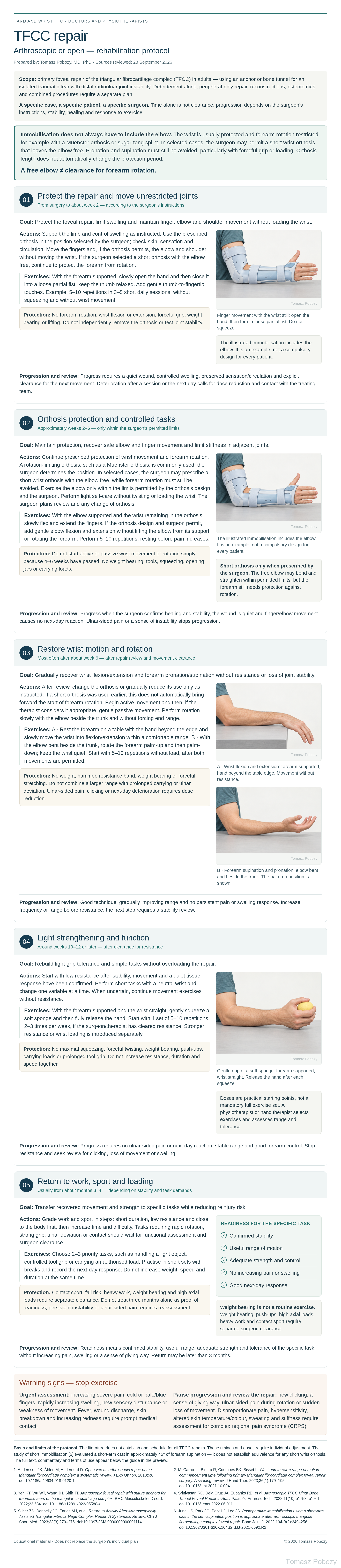
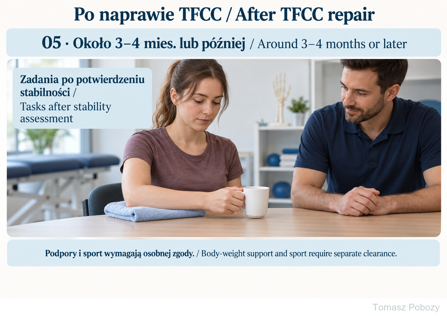
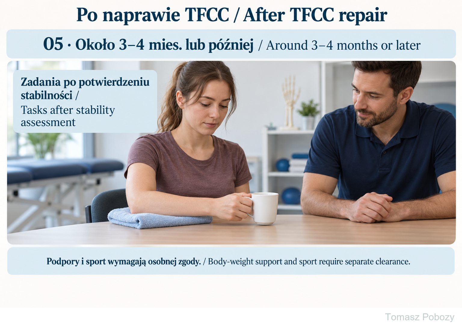
Phase 05: light grip and a specific task following stability confirmation, usually around months 3–4 or later. No fast twisting, body-weight support or forceful grip without separate clearance. Phases 01–04 on the full board show protection, continued finger motion, and later motion and resistance.
Exercises — a closer look
Use the complete boards above together with the instructions for the chosen stage. An exercise shown for a later stage is not permitted early.
Open the relevant complete board and zoom in. The viewer retains all stages and captions.
Exercise instructions and progression are contained in the stage descriptions linked above.
Phase 05: light grip and a specific task following stability confirmation, usually around months 3–4 or later. No fast twisting, body-weight support or forceful grip without separate clearance. Phases 01–04 on the full board show protection, continued finger motion, and later motion and resistance.
Scope and protection
Baseline assessment and operative details
Before starting, establish the exact repair variant (foveal, anchor or bone tunnel), arthroscopic or open approach, side, distal radioulnar joint stability, forearm position in the orthosis and all additional procedures. Record pain, swelling, wound, sensation, finger, elbow and shoulder movement, and after clearance also wrist and forearm rotation. Test grip strength only after resistance is permitted. Use the same measurements at follow-up and record the next-day response; weeks alone are not progression criteria. [1, 2, 3, 4, 5]
Immobilisation — rotation protection and a short orthosis
After foveal repair, immobilisation limiting wrist movement and forearm rotation is commonly used, for example a sugar-tong splint, a Muenster orthosis or another surgeon-selected solution. It may include the elbow or permit elbow flexion and extension while restricting forearm pronation and supination. The surgeon determines the type, position and duration of protection. The literature describes different protection periods; example techniques maintain protection for about 6 weeks, but this is not a universal prescription. [2, 4]
Short wrist orthosis — a possible exception. In selected cases, the surgeon may prescribe a short wrist orthosis that leaves the elbow free. This does not permit unrestricted forearm rotation. During the protective phase, pronation and supination must still be avoided, particularly with forceful gripping or loading; the surgeon specifies their permitted range and starting time. A free elbow does not mean unrestricted rotation.
The decision to use a short orthosis must consider the repair type, tissue quality, achieved stability and the patient’s ability to follow instructions. An exception concerning orthosis length does not automatically shorten the period required to protect the repaired tissues. Do not remove the orthosis or start forearm rotation, wrist flexion/extension or resistance without explicit clearance. For another repair variant, combined surgery, fracture or persistent instability, follow the surgeon’s individual plan.
A study of short immobilisation after foveal repair evaluated a short-arm cast in approximately 45° of forearm supination, not any removable wrist orthosis. It does not establish that all short orthoses are equivalent to immobilisation that includes the elbow. [6]
Stages and progression criteria
01 · Protect the repair and move unrestricted joints
From surgery to about week 2 — according to the surgeon’s instructions
Goal: Protect the foveal repair, limit swelling and maintain finger, elbow and shoulder movement without loading the wrist.
Actions: Support the limb and control swelling as instructed. Use the prescribed orthosis in the position selected by the surgeon; check skin, sensation and circulation. Move the fingers and, if the orthosis permits, the elbow and shoulder without moving the wrist. If the surgeon selected a short orthosis with the elbow free, continue to protect the forearm from rotation.
Exercises: With the forearm supported, slowly open the hand and then close it into a loose partial fist; keep the thumb relaxed. Add gentle thumb-to-fingertip touches. Example: 5–10 repetitions in 3–5 short daily sessions, without squeezing and without wrist movement.
Protection: No forearm rotation, wrist flexion or extension, forceful grip, weight bearing or lifting. Do not independently remove the orthosis or test joint stability.
Progression and review: Progress requires a quiet wound, controlled swelling, preserved sensation/circulation and explicit clearance for the next movement. Deterioration after a session or the next day calls for dose reduction and contact with the treating team.
02 · Orthosis protection and controlled tasks
Approximately weeks 2–6 — only within the surgeon’s permitted limits
Goal: Maintain protection, recover safe elbow and finger movement and limit stiffness in adjacent joints.
Actions: Continue prescribed protection of wrist movement and forearm rotation. A rotation-limiting orthosis, such as a Muenster orthosis, is commonly used; the surgeon determines the position. In selected cases, the surgeon may prescribe a short wrist orthosis with the elbow free, while forearm rotation must still be avoided. Exercise the elbow only within the limits permitted by the orthosis design and the surgeon. Perform light self-care without twisting or loading the wrist. The surgeon plans review and any change of orthosis.
Exercises: With the elbow supported and the wrist remaining in the orthosis, slowly flex and extend the fingers. If the orthosis design and surgeon permit, add gentle elbow flexion and extension without lifting the elbow from its support or rotating the forearm. Perform 5–10 repetitions, resting before pain increases.
Protection: Do not start active or passive wrist movement or rotation simply because 4–6 weeks have passed. No weight bearing, tools, squeezing, opening jars or carrying loads.
Progression and review: Progress when the surgeon confirms healing and stability, the wound is quiet and finger/elbow movement causes no next-day reaction. Ulnar-sided pain or a sense of instability stops progression.
03 · Restore wrist motion and rotation
Most often after about week 6 — after repair review and movement clearance
Goal: Gradually recover wrist flexion/extension and forearm pronation/supination without resistance or loss of joint stability.
Actions: After review, change the orthosis or gradually reduce its use only as instructed. If a short orthosis was used earlier, this does not automatically bring forward the start of forearm rotation. Begin active movement and then, if the therapist considers it appropriate, gentle passive movement. Perform rotation slowly with the elbow beside the trunk and without forcing end range.
Exercises: A · Rest the forearm on a table with the hand beyond the edge and slowly move the wrist into flexion/extension within a comfortable range. B · With the elbow bent beside the trunk, rotate the forearm palm-up and then palm-down; keep the wrist quiet. Start with 5–10 repetitions without load, after both movements are permitted.
Protection: No weight, hammer, resistance band, weight bearing or forceful stretching. Do not combine a larger range with prolonged carrying or ulnar deviation. Ulnar-sided pain, clicking or next-day deterioration requires dose reduction.
Progression and review: Good technique, gradually improving range and no persistent pain or swelling response. Increase frequency or range before resistance; the next step requires a stability review.
04 · Light strengthening and function
Around weeks 10–12 or later — after clearance for resistance
Goal: Rebuild light grip tolerance and simple tasks without overloading the repair.
Actions: Start with low resistance after stability, movement and a quiet tissue response have been confirmed. Perform short tasks with a neutral wrist and change one variable at a time. When uncertain, continue movement exercises without resistance.
Exercises: With the forearm supported and the wrist straight, gently squeeze a soft sponge and then fully release the hand. Start with 1 set of 5–10 repetitions, 2–3 times per week, if the surgeon/therapist has cleared resistance. Stronger resistance or wrist loading is introduced separately.
Protection: No maximal squeezing, forceful twisting, weight bearing, push-ups, carrying loads or prolonged tool grip. Do not increase resistance, duration and speed together.
Progression and review: Progress requires no ulnar-sided pain or next-day reaction, stable range and good forearm control. Stop resistance and seek review for clicking, loss of movement or swelling.
05 · Return to work, sport and loading
Usually from about months 3–4 — depending on stability and task demands
Goal: Transfer recovered movement and strength to specific tasks while reducing reinjury risk.
Actions: Grade work and sport in steps: short duration, low resistance and close to the body first, then increase time and difficulty. Tasks requiring rapid rotation, strong grip, ulnar deviation or contact should wait for functional assessment and surgeon clearance.
Exercises: Choose 2–3 priority tasks, such as handling a light object, controlled tool grip or carrying an authorised load. Practise in short sets with breaks and record the next-day response. Do not increase weight, speed and duration at the same time.
Protection: Contact sport, fall risk, heavy work, weight bearing and high axial loads require separate clearance. Do not treat three months alone as proof of readiness; persistent instability or ulnar-sided pain requires reassessment.
Progression and review: Readiness means confirmed stability, useful range, adequate strength and tolerance of the specific task without increasing pain, swelling or a sense of giving way. Return may be later than 3 months.
Warning signs
Warning signs — stop exercise and assess the cause
Urgent assessment: increasing severe pain, cold or pale/blue fingers, rapidly increasing swelling, new or worsening sensory disturbance or weakness of movement. Fever, wound discharge, skin breakdown and increasing redness require prompt medical contact. New clicking, a feeling that the joint is giving way, ulnar-sided pain with rotation or sudden loss of previously gained movement require suspension of progression and surgical review. Disproportionate pain, hypersensitivity, altered skin temperature or colour, sweating and progressive stiffness may indicate complex regional pain syndrome (CRPS) and require early assessment.
For a life-threatening emergency, call your local emergency number immediately — 112 in the European Union. On the boards, 112 refers to the EU.
Additional scope notes
Protecting the triangular fibrocartilage complex repair and gradually returning wrist motion, loading and function. The complete illustrated guide shows the stages and exercise technique; the expandable programme follows below.
This variant is for adults after primary arthroscopic or open foveal TFCC (triangular fibrocartilage complex) repair using a suture anchor or bone tunnel for an isolated traumatic tear with distal radioulnar joint instability. It does not cover isolated TFCC debridement without repair, peripheral-only repair, ligament reconstruction, extensor carpi ulnaris (ECU) subsheath repair, ulnar shortening, simultaneous fracture fixation or other combined procedures; these variants require separate surgeon-defined restrictions.
A specific case, a specific patient, a specific surgeon. Timings are reference points, not automatic permissions. Operative instructions, distal radioulnar joint stability, wound status, pain, movement and surgical review determine progression. If a different repair variant was performed, this schedule does not replace the orthopaedic surgeon’s plan.
Doses of 5–10 slow repetitions, 3–5 short daily sessions and 2–3 strengthening sessions per week are practical starting points for individual adjustment. Reviews describe widely varying protocols and do not establish one universal movement schedule; do not perform the entire set without selection by the treating team.
Short wrist orthosis — a possible exception. In selected cases, the surgeon may prescribe a short wrist orthosis that leaves the elbow free. This does not permit unrestricted forearm rotation. During the protective phase, pronation and supination must still be avoided, particularly with forceful gripping or loading; the surgeon specifies their permitted range and starting time. A free elbow does not mean unrestricted rotation.
References
References
- Andersson JK, Åhlén M, Andernord D. Open versus arthroscopic repair of the triangular fibrocartilage complex: a systematic review. J Exp Orthop. 2018;5:6. doi:10.1186/s40634-018-0120-1
- McCarron L, Bindra R, Coombes BK, Bisset L. Wrist and forearm range of motion commencement time following primary triangular fibrocartilage complex foveal repair surgery: A scoping review. J Hand Ther. 2023;36(1):179–195. doi:10.1016/j.jht.2021.10.004
- Yeh KT, Wu WT, Wang JH, Shih JT. Arthroscopic foveal repair with suture anchors for traumatic tears of the triangular fibrocartilage complex. BMC Musculoskelet Disord. 2022;23:634. doi:10.1186/s12891-022-05588-z
- Srinivasan RC, Dela Cruz JA, Eubanks RD, et al. Arthroscopic TFCC Ulnar Bone Tunnel Foveal Repair in Adult Patients. Arthrosc Tech. 2022;11(10):e1753–e1761. doi:10.1016/j.eats.2022.06.011
- Silber ZS, Donnelly JC, Farias MJ, et al. Return to Activity After Arthroscopically Assisted Triangular Fibrocartilage Complex Repair: A Systematic Review. Clin J Sport Med. 2023;33(3):270–275. doi:10.1097/JSM.0000000000001114
- Jung HS, Park JG, Park HJ, Lee JS. Postoperative immobilization using a short-arm cast in the semisupination position is appropriate after arthroscopic triangular fibrocartilage complex foveal repair. Bone Joint J. 2022;104-B(2):249–256. doi:10.1302/0301-620X.104B2.BJJ-2021-0592.R2
References and links checked on 18 October 2026. Existence of a publication does not validate every dose or timetable. Institutional protocols and examples require individualisation.
Clinical commentary and terms of use
Commentary — repair and rehabilitation are one process
The arthroscopic or open approach alone does not automatically determine the pace of rehabilitation. The repaired structures, fixation method, tissue quality and achieved stability matter. This schedule remains a foveal-repair pathway; it should not be applied unchanged to debridement alone or to every type of peripheral repair. [1, 2, 3, 4]
A foveal repair is intended to restore ligamentous stability, but early repair strength is not the same as full load tolerance. The physiotherapist or hand therapist assesses swelling, movement, technique and post-session response; the surgeon sets restrictions arising from the suture, tunnel, tissue tension and joint examination. Feedback should include ulnar-sided pain, clicking, a sense of instability and function, not range alone. Do not force a stiff wrist or advance rotation simply because the wound looks healed.
Systematic and scoping reviews report improved outcomes after TFCC repair but also substantial variation in techniques and no single evidence-based rehabilitation protocol. Studies and technique reports describe wrist movement starting around weeks 2–8, forearm rotation around weeks 2–12, and strengthening around week 10 or later. One retrospective foveal-repair study used 6 weeks of immobilisation, then movement and isometrics, with progression to strengthening after another month; this does not prove a mandatory schedule. Return to sport/work is often reported around 3 months, but the exact timing depends on stability, function and task demands. [1, 2, 3, 4, 5]