 Skip to content
FOR CLINICIANS AND PHYSIOTHERAPISTS
Achilles tendon repair — rehabilitation protocol
Protecting the repair from excessive elongation, progressive weight bearing, controlled restoration of motion and rebuilding calf strength. The timetable is a clinical framework rather than an automatic prescription.
Using and loading the limb
Use the surgeon-selected loading pathway: initially NWB, partial or progressive protected loading may apply. Absence of pain does not permit acceleration.
Full guidance ↓Brace or immobilisation
Use a boot with wedges or other prescribed immobilisation in plantarflexion. Remove wedges in stages according to the plan.
Full guidance ↓Permitted movement
Protect against tendon elongation. During weeks 2–6, move toward neutral 0° without dorsiflexing beyond it unless specifically prescribed.
Full guidance ↓Permitted movement, loading and removal of a brace are separate decisions. Additional repairs may require a more protective programme.
Rehabilitation at a glance
Progression follows healing and functional criteria, not dates alone.
Rehabilitation boards
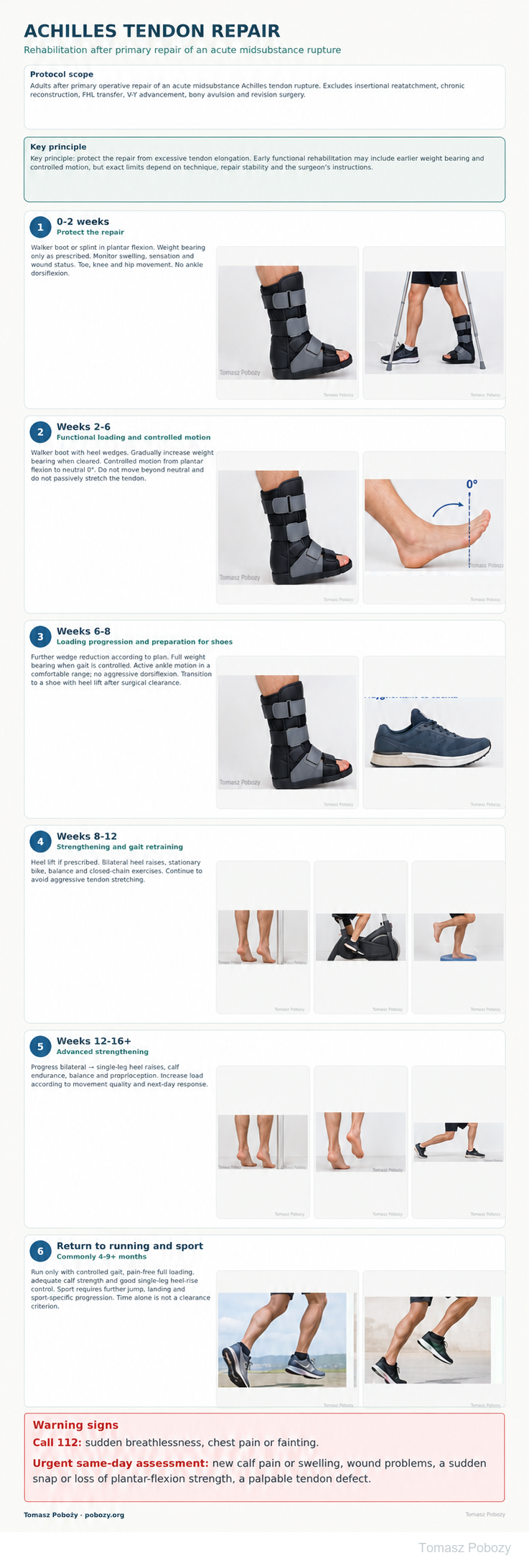
Exercises — a closer look
Use the complete boards above together with the instructions for the chosen stage. An exercise shown for a later stage is not permitted early.
Open the relevant complete board and zoom in. The viewer retains all stages and captions.
Exercise instructions and progression are contained in the stage descriptions linked above.
Scope and protection
Baseline assessment and operative details
Before starting, establish rupture location, repair technique, the protective ankle position, weight-bearing instructions, the prescribed boot or immobilisation, associated procedures and the surgical follow-up plan. Assess sensation, perfusion, swelling, wound status, knee and hip movement, and safe mobility with an aid.
For insertional repairs, chronic reconstruction, tendon transfers, bony avulsion or revision surgery, follow the surgeon-specific programme rather than automatically applying this schedule.
Return to running and sport
Return to running is criteria-based rather than calendar-based. Requirements include pain-free full weight bearing, controlled gait, good heel-rise tolerance and adequate calf strength and endurance. In practice, running and sport usually return after several months, commonly around 4–9 months or later depending on the sport and function.
Before sports involving running, cutting and jumping, introduce progressive plyometrics, controlled landing and sport-specific tasks. No single universal return-to-sport test clears every patient.
Stages and progression criteria
Stage I — weeks 0–2: protect the repair
A walker boot with heel wedges or another prescribed immobilisation maintains the ankle in plantar flexion. Weight bearing is according to the surgeon’s instructions and may be partial or progressively increased depending on the repair.
Monitor swelling and walking tolerance. Maintain toe, knee and hip movement when permitted. Do not dorsiflex the ankle or passively stretch the repaired tendon.
Stage II — weeks 2–6: functional loading and controlled motion
Continue the walker with heel wedges. Gradually increase weight bearing when cleared and when gait remains controlled. Wedge reduction is gradual and follows the treating team’s plan.
Controlled ankle motion progresses from plantar flexion towards neutral. Do not move beyond neutral 0° into dorsiflexion during this period unless the surgeon specifically permits it. Avoid aggressive calf stretching and forced dorsiflexion.
Stage III — weeks 6–8: loading progression and preparation for shoes
Continue gradual wedge reduction and progress towards full weight bearing if gait is controlled, pain and swelling are acceptable and healing is satisfactory. Introduce active ankle motion within a comfortable range, still avoiding aggressive end-range dorsiflexion.
Transition to a normal shoe may temporarily require a heel lift. Timing and heel-lift height depend on surgical review and gait quality.
Stage IV — weeks 8–12: strengthening and gait retraining
Develop a controlled gait pattern, calf strength and lower-limb stability. The programme can include supported bilateral heel raises, stationary cycling, balance exercises and closed-chain tasks.
Increase load gradually. Absence of pain during an exercise is not, by itself, a reason to rapidly increase dorsiflexion range or stretching intensity.
Stage V — weeks 12–16+: advanced strengthening
Progress from bilateral towards single-leg heel raises, improve calf endurance, balance and proprioception, and introduce progressively more demanding functional tasks. Increase repetitions, range and external load separately while observing the immediate and next-day response.
Persistent marked heel-rise weakness, increasing swelling or deterioration in gait quality requires programme modification and reassessment.
Warning signs
Call 112: sudden breathlessness, chest pain or fainting. New calf pain/swelling, wound breakdown, cold or blue toes or sudden loss of function require urgent same-day assessment.
Warning signs — stop progression and reassess
Urgent review is required for sudden loss of plantar-flexion strength, a new palpable tendon defect, a snap/pop sensation or abrupt functional deterioration. Increasing pain and swelling, wound-healing problems, fever or wound discharge also require medical review.
New calf pain and swelling requires assessment for deep-vein thrombosis. Sudden shortness of breath or chest pain requires emergency assessment.
For a life-threatening emergency, call your local emergency number immediately — 112 in the European Union. On the boards, 112 refers to the EU.
Additional scope notes
FOOT · ACHILLES TENDON · FOR DOCTORS AND PHYSIOTHERAPISTS
Prepared by: Tomasz Poboży, MD, PhD — orthopaedic surgeon
Sources reviewed: 28 September 2026
Scope. This protocol is for adults after primary operative repair of an acute midsubstance Achilles tendon rupture. It does not automatically apply to insertional reattachment at the calcaneus, chronic reconstruction, flexor hallucis longus (FHL) transfer, V-Y advancement, bony avulsion or revision surgery.
The operative record, repair stability and the orthopaedic surgeon’s instructions take priority over the timetable on this page.
Everyday activities
Functional clearance takes priority over the calendar.
Walking and stairs
Keep the prescribed loading limit and protection. Practise transfers and stairs with a physiotherapist; use a handrail and assistance until the technique is safe.
Driving and work
Driving requires safe emergency braking, no immobilisation interfering with the pedals and no medication impairing alertness. Operated side and transmission matter. Desk work requires safe access and the ability to elevate the leg; standing work depends on loading clearance and tolerance.
References
References
- Massen FK, et al. Rehabilitation following operative treatment of acute Achilles tendon ruptures: a systematic review and meta-analysis. EFORT Open Rev. 2022;7(10):680–691. doi:10.1530/EOR-22-0072.
- Huang J, Wang C, Ma X, et al. Rehabilitation regimen after surgical treatment of acute Achilles tendon ruptures: a systematic review with meta-analysis. Am J Sports Med. 2015;43(4):1008–1016. doi:10.1177/0363546514531014.
- McCormack R, Bovard J. Early functional rehabilitation or cast immobilisation for postoperative management of acute Achilles tendon rupture? Br J Sports Med. 2015;49(20):1329–1335. doi:10.1136/bjsports-2015-094935.
- Gould HP, Bano JM, Akman JL, Fillar AL. Postoperative Rehabilitation Following Achilles Tendon Repair: A Systematic Review. Sports Med Arthrosc Rev. 2021. doi:10.1097/JSA.0000000000000309.
- Valkering KP, et al. Functional weight-bearing mobilization after Achilles tendon rupture enhances early healing response: a single-blinded randomized controlled trial. Knee Surg Sports Traumatol Arthrosc. 2017;25:1807–1816. doi:10.1007/s00167-016-4270-3.
- Costa ML, et al. Randomised controlled trials of immediate weight-bearing mobilisation for rupture of the tendo Achillis. J Bone Joint Surg Br. 2006;88(1):69–77. doi:10.1302/0301-620X.88B1.16549.
- Čretnik A, Košir R. Prospective randomized comparison of functional bracing versus rigid immobilization with early weightbearing after modified percutaneous Achilles tendon repair under local anesthesia. Foot (Edinb). 2024;60:102124. doi:10.1016/j.foot.2024.102124.
Clinical commentary and terms of use
Commentary — functional rehabilitation still requires protection
Randomized trials and systematic reviews support earlier functional weight bearing and controlled motion after stable operative repair of an acute Achilles rupture. This does not mean unrestricted dorsiflexion or free stretching during the first weeks. Published protocols vary in weight-bearing timing, range of motion and wedge reduction.1–7
Limiting excessive tendon elongation remains a major early goal because elongation can impair later calf strength and heel-rise function. The surgeon communicates repair-specific protection limits while the physiotherapist individualizes loading, exercise selection and progression criteria according to current function.