 Przejdź do treści
FOR CLINICIANS AND PHYSIOTHERAPISTS
Intertrochanteric femoral fracture — Gamma nail fixation — rehabilitation protocol
Protection, movement and a gradual return to daily activity. Select the stage appropriate to the actual procedure and current clearance.
Using and loading the limb
After stable fixation, aim for early mobilisation and walking with aids. Exact loading depends on fracture, reduction quality and prescription.
Full guidance ↓Brace or immobilisation
This programme does not prescribe a routine hip brace. Additional protection needs a specific indication; avoid unnecessary immobilisation.
Full guidance ↓Permitted movement
Use early permitted gentle hip, knee and ankle motion. Avoid painful forcing, pivoting on a planted foot and resistance ahead of healing.
Full guidance ↓Permitted movement, loading and removal of a brace are separate decisions. Additional repairs may require a more protective programme.
Rehabilitation at a glance
Progression follows healing and functional criteria, not dates alone.
Rehabilitation boards
Exercises — a closer look
Use the complete boards above together with the instructions for the chosen stage. An exercise shown for a later stage is not permitted early.
Open the relevant complete board and zoom in. The viewer retains all stages and captions.
Exercise instructions and progression are contained in the stage descriptions linked above.
Scope and protection
Prevention of future fractures
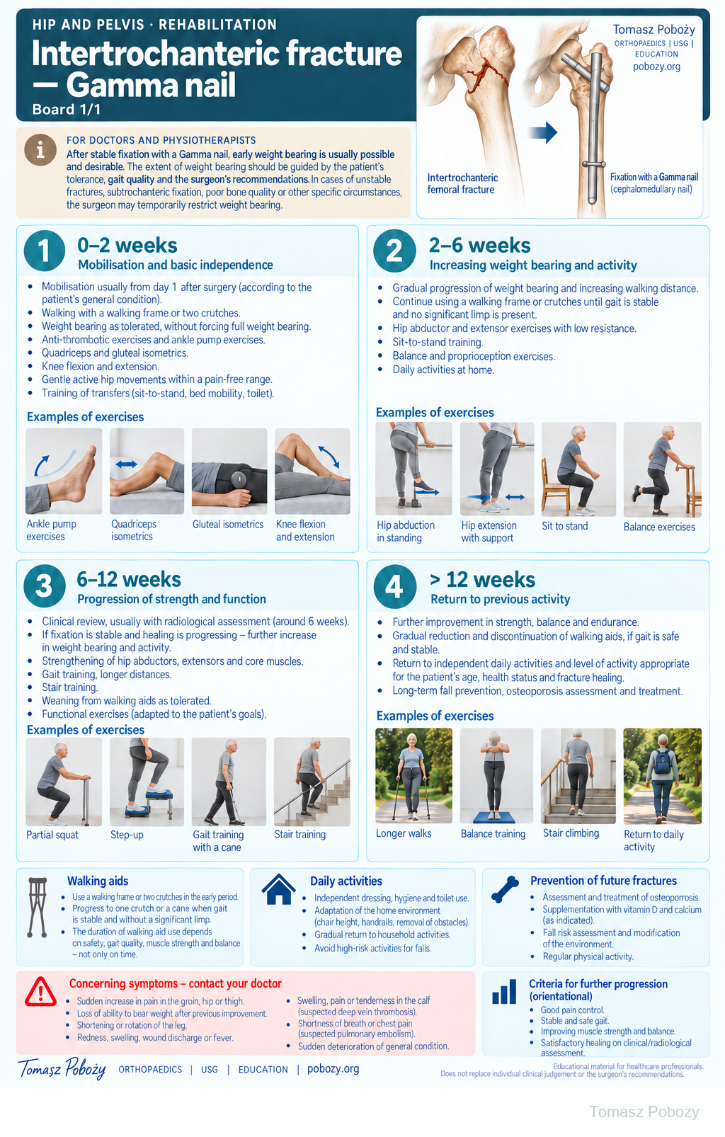
After a typical low-energy proximal femoral fracture, assess osteoporosis and future fall risk. Further management may include osteoporosis investigation and treatment, vitamin D and calcium when indicated, environmental modification and regular physical activity appropriate to the patient’s abilities.
Stages and progression criteria
Phase 1 · 0–2 weeks — mobilisation and basic independence
Mobilisation usually starts on postoperative day 1, depending on general condition. Walk with a frame or two crutches. Increase weight bearing on the operated limb according to pain tolerance and the orthopaedic surgeon’s instructions, without forcing full loading. Exercises include ankle pumps, quadriceps and gluteal isometrics, knee flexion and extension, and gentle active hip movement. Practise safe transfers, including sit-to-stand, bed mobility and toilet use.
Phase 2 · 2–6 weeks — increasing weight bearing and activity
Gradually increase weight bearing and walking distance. Continue using a walking aid while gait remains uncertain or there is a clear limp. Introduce low-resistance hip abductor and extensor work, sit-to-stand training, balance and trunk-stability exercises. Increase daily activity according to function rather than the calendar alone.
Phase 3 · 6–12 weeks — progression of strength and function
Clinical review is usually performed at around 6 weeks, often with radiological assessment. If fixation is stable and healing is progressing appropriately, further increase weight bearing and activity. Strengthen the gluteal, thigh and core muscles; add shallow squats, step-ups, longer walks, gait training and stair practice. Reduce walking aids only when gait remains safe and stable.
Phase 4 · >12 weeks — return to activity
Continue improving strength, balance and endurance. Gradually reduce and discontinue walking aids if gait is safe and stable. Aim for the highest feasible level of independence and return to pre-fracture activities according to age, general health, previous function and fracture healing.
Daily activities and follow-up
Walking aids and daily activities
- A walking frame or two crutches are usually appropriate early on.
- Progress to one crutch or a cane only when gait is stable without a marked limp.
- The duration of walking-aid use depends on safety, gait quality, strength and balance — not simply on time.
- Practise independent dressing, hygiene and toilet use; adapt chair height, handrails and the home environment to reduce fall risk.
Warning signs
Warning signs — medical reassessment
Seek prompt reassessment for a sudden increase in groin, hip or thigh pain; loss of weight-bearing ability after previous improvement; shortening or rotation of the limb; increasing redness, swelling, wound discharge or fever; calf pain or swelling; shortness of breath or chest pain; or sudden deterioration in function or general condition.
Further guidance
HIP AND PELVIS · FOR DOCTORS AND PHYSIOTHERAPISTS
Early mobilisation, graded weight bearing, restoration of safe gait and independence after fixation with a Gamma nail (cephalomedullary intramedullary nail).
Prepared by: Tomasz Poboży, MD, PhD · Updated: 03 October 2026
Scope
This protocol applies to adults after operative fixation of an intertrochanteric femoral fracture with a Gamma nail. Weight-bearing and progression depend on fracture pattern, bone quality, reduction, fixation stability, general condition and the surgeon’s individual instructions.
Key principle: after stable fixation, early weight bearing is usually possible and desirable. Its extent should increase according to the patient’s tolerance, gait quality and the surgeon’s recommendations. Unstable fractures, suboptimal fixation stability, poor bone quality or other specific circumstances may require temporary restriction.
Key message: rehabilitation aims for an early, safe return to walking and independence without unnecessary immobilisation, while respecting fixation stability and individual patient limitations.
References
References and basis of recommendations
- American Academy of Orthopaedic Surgeons. Management of Hip Fractures in Older Adults. Clinical Practice Guideline. 2021.
- National Institute for Health and Care Excellence. Hip fracture: management. Clinical guideline CG124. Updated recommendations.
- Warren J, Sundaram K, Anis H, et al. The association between weight-bearing status and early complications in hip fractures. Eur J Orthop Surg Traumatol. 2019;29:1419–1427.
Guidelines support early mobilisation and, after sufficiently stable fixation, early weight bearing as tolerated. They do not replace the surgeon’s assessment of the individual fracture and fixation construct.