 Przejdź do treści
FOR CLINICIANS AND PHYSIOTHERAPISTS
Femoral neck fracture — cannulated screw fixation — rehabilitation protocol
Protection, movement and a gradual return to daily activity. Select the stage appropriate to the actual procedure and current clearance.
Using and loading the limb
There is no universal full-loading date after screw fixation. Older patients may be permitted early WBAT; younger patients often need initial protection and review.
Full guidance ↓Brace or immobilisation
Do not prescribe a routine brace from the procedure name alone. Walking aids and the loading limit are prescribed individually.
Full guidance ↓Permitted movement
Use gentle permitted hip, knee and ankle movement without painful forcing. Restored motion does not prove union or remove walking limits.
Full guidance ↓Permitted movement, loading and removal of a brace are separate decisions. Additional repairs may require a more protective programme.
Rehabilitation at a glance
Progression follows healing and functional criteria, not dates alone.
Rehabilitation boards
Exercises — a closer look
Use the complete boards above together with the instructions for the chosen stage. An exercise shown for a later stage is not permitted early.
Open the relevant complete board and zoom in. The viewer retains all stages and captions.
Exercise instructions and progression are contained in the stage descriptions linked above.
Scope and protection
When to suspect impaired healing?
Persistent or increasing pain or absent healing progression on serial imaging requires reassessment. Do not wait until 6 months if the course is concerning. Imaging, including CT when indicated, and treatment are selected by the orthopaedic surgeon.
Stages and progression criteria
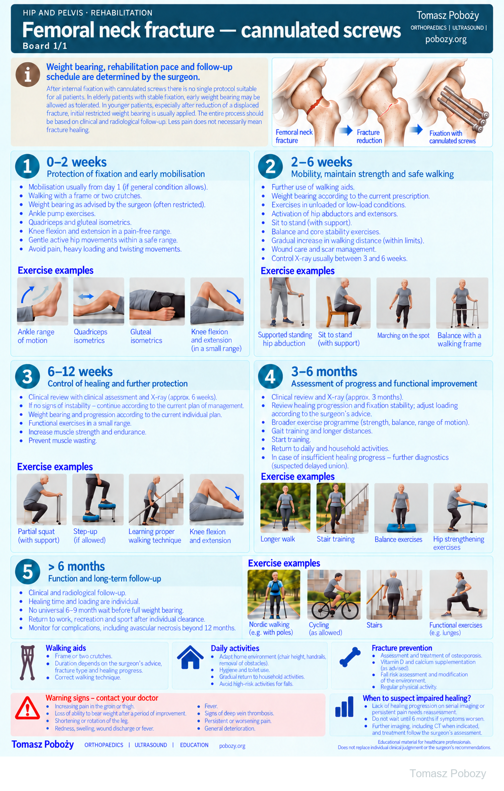
0–2 weeks — early mobilisation and an individual loading plan
Mobilisation usually starts from postoperative day 1 if general condition allows. Walk with a frame or two crutches. Confirm the permitted weight-bearing status before walking. In older patients, weight bearing as tolerated may be permitted early after appropriate fixation; younger patients may require initial restriction with reassessment at approximately 6 weeks. Do not assume that the same restriction applies to everyone. AO Surgery Reference; NICE CG124.
Use ankle pumps, quadriceps and gluteal isometrics, knee flexion and extension, and gentle pain-free hip movements. Avoid painful loading, falls, twisting movements and heavy resistance.
2–6 weeks — mobility, maintenance of strength and safe walking
Continue walking aids and the prescribed weight-bearing status. Use unloaded or low-resistance exercises, activate hip abductors and extensors, practise supported sit-to-stand, balance and core stability. Control X-ray between approximately 3 and 6 weeks is primarily used to assess fracture position and fixation stability; complete union is not expected at this stage.
6–12 weeks — fixation follow-up and continued protection
Clinical and radiological review is usually performed around 6 weeks. If there is no evidence of instability, continue management according to the surgeon’s instructions. Weight bearing may still be restricted. Functional exercises should remain within a safe range without excessive loading. Maintain trunk and limb strength and practise correct gait while preventing muscle wasting.
3–6 months — healing review and functional progression
Clinical and radiological review assesses the course of healing and the stability of fixation. Do not interpret this period as the earliest possible appearance of healing or a universal date for starting full weight bearing. Progress strength, balance, range of motion and walking according to the surgeon’s current plan and clinical findings. Persistent or increasing pain and lack of healing progression require reassessment.
Beyond 6 months — function and long-term surveillance
Continue rehabilitation according to remaining functional deficits. There is no universal requirement to wait 6–9 months before full weight bearing after cannulated-screw fixation. Return to work, recreation and sport depends on healing, function and individual clearance. Follow-up also considers avascular necrosis of the femoral head, including beyond 12 months.
Daily activities and follow-up
Fracture prevention and long-term follow-up
When appropriate, assess and treat osteoporosis, fall risk, and vitamin D or calcium deficiency. Encourage regular physical activity adapted to the patient’s abilities. Long-term follow-up includes hip function, fracture union and surveillance for possible avascular necrosis of the femoral head.
Warning signs
Warning signs
New severe pain, sudden inability to bear the previously permitted load, a fall with functional deterioration, wound discharge or fever require prompt medical assessment. Sudden shortness of breath, chest pain or collapse require emergency help.
Further guidance
HIP AND PELVIS · FOR DOCTORS AND PHYSIOTHERAPISTS
Protection of fixation, safe mobilisation and gradual functional progression while accounting for the relatively slow healing of femoral neck fractures.
Prepared by: Tomasz Poboży, MD, PhD · Updated: 03 October 2026
Key principle
Weight bearing, rehabilitation pace and follow-up imaging are determined by the surgeon. There is no single protocol suitable for every patient after cannulated-screw fixation. In elderly patients with stable fixation, earlier weight bearing as tolerated may be appropriate. In younger patients, particularly after reduction of a displaced fracture, initial restricted weight bearing is usually used. Progression should be based on clinical and radiological follow-up. Less pain does not necessarily mean fracture union.
Key message: after femoral neck fixation, the calendar does not replace clinical and radiological assessment. Weight-bearing progression should reflect fixation quality and actual healing progress.
References
References
- AO Foundation Surgery Reference. Femoral neck fracture — cannulated screws: aftercare and weight bearing.
- American Academy of Orthopaedic Surgeons. Management of Hip Fractures in Older Adults. Clinical Practice Guideline. 2021.
- National Institute for Health and Care Excellence. Hip fracture: management. CG124.