 Skip to content
FOOT AND ANKLE / POSTOPERATIVE PROTOCOL
Fifth metatarsal fracture — postoperative rehabilitation
After surgery for a fifth metatarsal fracture. The main pathway addresses fixation of a Jones or proximal shaft fracture; avulsion fractures and nonunion revision require the modifications described below.
For clinicians, physiotherapists and patients. The individual plan based on the operative report takes precedence over the time ranges on this page.
Essential protection
- Weight-bearing
- After Jones fracture fixation, an initial non-weight-bearing period of around 4 weeks is commonly followed by cautious progression in a boot. Low pain alone does not justify increasing load; healing must be assessed.
- Brace / immobilisation
- Initial splint/cast, then a walking boot as prescribed.
- Range of motion
- Ankle motion after clearance; avoid forced adduction/inversion and early resisted eversion when the tendon insertion has been repaired.
Rehabilitation at a glance
Timings are approximate. Progression requires criteria and clearance for the relevant motion or loading level.
- 01
Protecting fixation
Weeks 0–4Protect the wound and a fracture site at increased risk of healing problems.
- 02
Movement and cautious loading
Usually weeks 4–6Restore gentle movement and prepare for walking.
- 03
Walking after healing review
Usually weeks 6–12Reduce crutch and boot use without recurrent pain.
- 04
Control and return to sport
From around week 12; criteria basedReturn to activity while reducing refracture risk.
Full rehabilitation board
Click to enlarge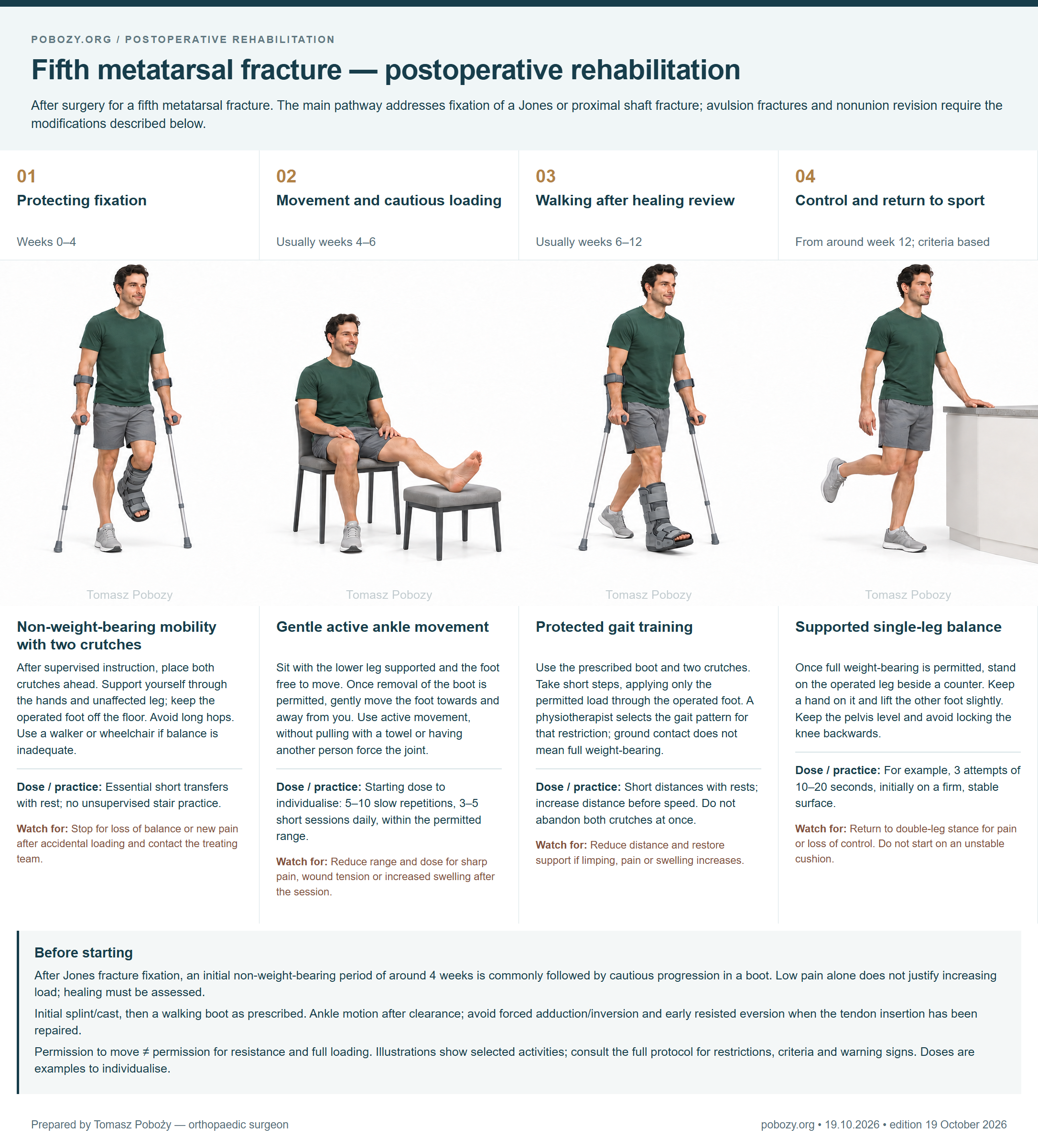
Exercises — take a closer look4 phases • technique • dosage • when to stop
Each phase has its own illustration. The descriptions below are the text equivalent of the board. Doses are starting examples to individualise with a physiotherapist and do not override surgical restrictions.
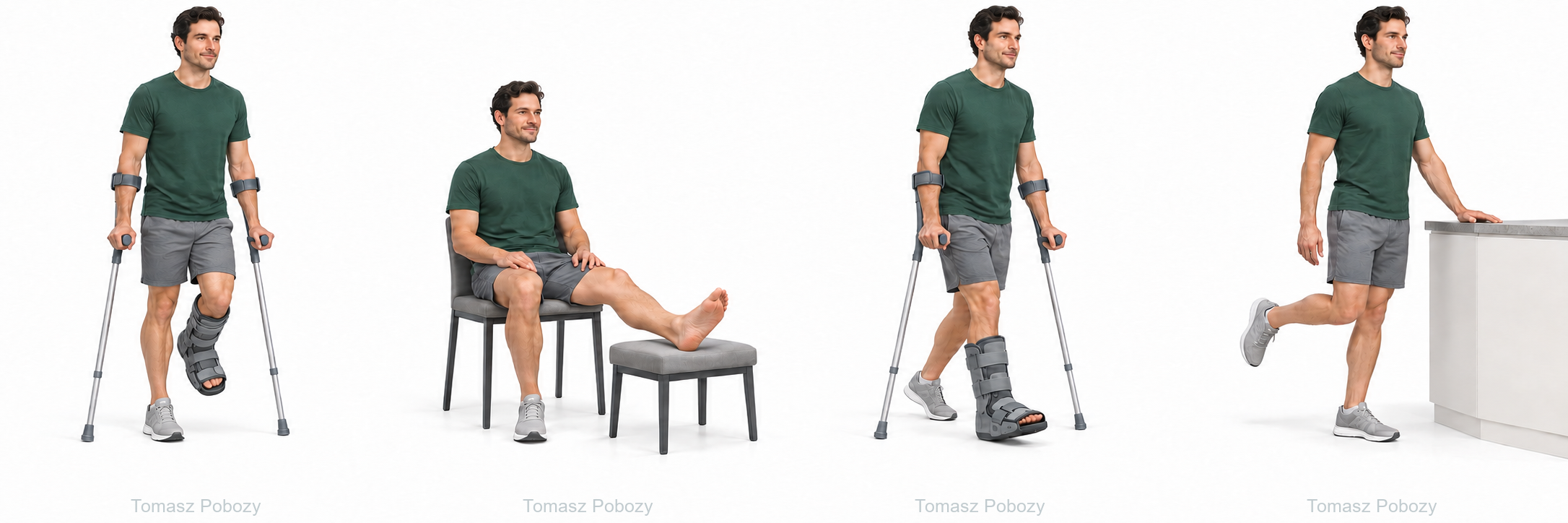
Non-weight-bearing mobility with two crutches
After supervised instruction, place both crutches ahead. Support yourself through the hands and unaffected leg; keep the operated foot off the floor. Avoid long hops. Use a walker or wheelchair if balance is inadequate.
- Dose to individualise
- Essential short transfers with rest; no unsupervised stair practice.
- When to reduce or stop
- Stop for loss of balance or new pain after accidental loading and contact the treating team.
Gentle active ankle movement
Sit with the lower leg supported and the foot free to move. Once removal of the boot is permitted, gently move the foot towards and away from you. Use active movement, without pulling with a towel or having another person force the joint.
- Dose to individualise
- Starting dose to individualise: 5–10 slow repetitions, 3–5 short sessions daily, within the permitted range.
- When to reduce or stop
- Reduce range and dose for sharp pain, wound tension or increased swelling after the session.
Protected gait training
Use the prescribed boot and two crutches. Take short steps, applying only the permitted load through the operated foot. A physiotherapist selects the gait pattern for that restriction; ground contact does not mean full weight-bearing.
- Dose to individualise
- Short distances with rests; increase distance before speed. Do not abandon both crutches at once.
- When to reduce or stop
- Reduce distance and restore support if limping, pain or swelling increases.
Supported single-leg balance
Once full weight-bearing is permitted, stand on the operated leg beside a counter. Keep a hand on it and lift the other foot slightly. Keep the pelvis level and avoid locking the knee backwards.
- Dose to individualise
- For example, 3 attempts of 10–20 seconds, initially on a firm, stable surface.
- When to reduce or stop
- Return to double-leg stance for pain or loss of control. Do not start on an unstable cushion.
Who this protocol is for
After surgery for a fifth metatarsal fracture. The main pathway addresses fixation of a Jones or proximal shaft fracture; avulsion fractures and nonunion revision require the modifications described below.
Excludes nonoperative treatment and Lisfranc injury. Revision, bone grafting or peroneus brevis repair must be reflected in an individual plan.
Before starting, confirm the operation date and extent, fixation, permitted loading, support-removal rules, range of motion and review date. Obtain any missing instruction from the surgeon before progressing.
Full programme by phase
01 · Protecting fixationWeeks 0–4
- Goal
- Protect the wound and a fracture site at increased risk of healing problems.
- Weight-bearing
- Typical Jones pathway: initially non-weight-bearing. Earlier loading is an alternative only when specifically authorised by the surgeon.
- Support / immobilisation
- Splint or cast, followed by a boot according to the plan.
- Motion
- Move the hip, knee and unrestricted toes; ankle motion requires permission to remove the support.
- Work in this phase
- Safe non-weight-bearing crutch mobility, swelling control and thigh activation.
- Progression criteria
- Wound healing, maintained fixation and clearance for the next level of protection or movement.
02 · Movement and cautious loadingUsually weeks 4–6
- Goal
- Restore gentle movement and prepare for walking.
- Weight-bearing
- Partial weight-bearing in a boot only when cleared; zone 3 injuries, revision or poor healing progress may require longer protection.
- Support / immobilisation
- Boot for permitted walking; removal for exercise follows the treating team’s instructions.
- Motion
- Active dorsiflexion and plantarflexion without lateral-foot pain. Avoid sudden twisting.
- Work in this phase
- Short movement sessions; no heel raises, running, hopping or resisted eversion without clearance.
- Progression criteria
- Review around week 6 or as scheduled: assess tenderness and imaging before further loading.
03 · Walking after healing reviewUsually weeks 6–12
- Goal
- Reduce crutch and boot use without recurrent pain.
- Weight-bearing
- Full weight-bearing and footwear transition require adequate union and clearance. Lack of union means revising the plan, not proceeding by the calendar.
- Support / immobilisation
- Supportive shoes once cleared; avoid deliberate loading on the inverted outer border of the foot.
- Motion
- Gradually restore motion and light resistance after assessment of bone and any tendon repair.
- Work in this phase
- Gait training and double-leg loading tasks within the permitted dose; avoid premature impact exercise.
- Progression criteria
- No fracture-site tenderness, normal gait, radiographic healing progress and clearance for greater demands.
04 · Control and return to sportFrom around week 12; criteria based
- Goal
- Return to activity while reducing refracture risk.
- Weight-bearing
- Running and direction changes require confirmed union and strength assessment. In elite athletes or uncertain union, the clinician may request CT.
- Support / immobilisation
- Activity-appropriate footwear; assess alignment when overload recurs.
- Motion
- Task-appropriate movement without lateral-foot pain.
- Work in this phase
- Balance on a stable surface, calf and foot strength, then graded sport loading.
- Progression criteria
- Union, no pain, sport-appropriate capacity and no next-day deterioration.
How to adjust the dose
Increase one element at a time: repetitions, distance, range or resistance. Sharp pain, limping or markedly increased swelling calls for reducing the load. Check symptoms after exercise and the following day; discuss persistent deterioration. Absence of pain does not replace union assessment.
If the surgeon prescribes a kilogram limit, record the value and duration. Do not independently convert “as tolerated” into kilograms. Do not add weights without clearance for resistance.
Everyday activities and return to function
- Walking and stairs
- Use aids while needed to respect the load restriction and maintain gait quality. Practise stairs with a physiotherapist; choose a safer alternative when support or a rail is unavailable.
- Driving
- Resume only after assessment of rapid, forceful emergency braking and without medication impairing alertness. Operated side, transmission, bracing and loading restrictions affect the decision; discuss it with the treating team and consider insurance conditions.
- Work, rest and swelling
- Seated work requires opportunities to change position and elevate the leg; standing or manual work requires loading and task tolerance. Swelling may persist, but new or rapidly increasing swelling requires assessment.
- Wound care and prevention
- Do not soak an unhealed wound, remove wires yourself or change prescribed thromboprophylaxis. Follow individual medication, dressing, compression and scar-care instructions. Swimming requires complete wound closure and clearance.
- Running and sport
- Timing follows healing and capacity; it is not a stand-alone criterion. Establish safe gait, strength, balance and task tolerance first, progressing without significant next-day worsening.
Warning signs — when to seek help
- Emergency help
- Sudden shortness of breath, chest pain, collapse or coughing up blood: call emergency services. Use 112 in the EU; elsewhere use the appropriate local emergency number.
- Urgent same-day assessment
- New one-sided calf pain and swelling; increasing pain despite rest; fever with wound redness or discharge; wound breakdown; a new injury or pain after accidental loading.
- Do not wait for a routine appointment
- A cold, pale or blue foot, rapidly increasing numbness, loss of toe movement or severe increasing pain under a cast requires immediate emergency assessment.
Variants and clinical context
- Zone 1: tuberosity avulsion, usually treated nonoperatively. After surgery, protection depends on the fragment and any tendon insertion repair. Do not automatically apply the Jones pathway.
- Zone 2: a Jones fracture at the metaphyseal–diaphyseal junction extending into the fourth–fifth intermetatarsal articulation. Healing risk is higher than with a typical avulsion fracture.
- Zone 3: a proximal shaft fracture, often stress related. Delayed union, nonunion and revision require individually extended protection and risk-factor assessment.
- A distal shaft or neck fracture of the fifth metatarsal is not automatically a Jones fracture. Establish classification from imaging and the operative report.
Related resources
References and evidenceVerified publications and evidence limitations
- Fifth metatarsal fractures: an update on management, complications, and outcomes. EFORT Open Reviews. 2022. DOI: 10.1530/EOR-21-0025.
- Republication of “Metatarsal Fractures”. Foot & Ankle Orthopaedics. 2023. DOI: 10.1177/24730114231193428.
Research informs protection and progression decisions; it does not validate every illustrated exercise or example dose. Procedure details, tissue condition and the individual plan determine application.
Glossary
- ROM
- Joint range of motion. Active movement uses your own muscles; assisted movement uses additional help.
- Non-weight-bearing
- No body-weight transfer through the operated foot during standing and walking.
- As tolerated
- Once this pathway is authorised: loading limited by symptoms and movement quality within prescribed protection. It is not permission for unrestricted sport.
- Walker
- A walking boot is a protective ankle-foot support. A walking frame is a separate mobility aid.