 Skip to content
FOR CLINICIANS AND PHYSIOTHERAPISTS
Finger flexor tendon repair — rehabilitation protocol
Protecting the repair, restoring controlled tendon glide and gradually returning function. The protocol includes several early strategies—from temporary immobilisation, through Kleinert and Duran, to early active motion—with particular emphasis on Zone II repairs.
Using and loading the limb
Early therapeutic movement does not permit gripping or carrying objects. Do not independently test repair strength.
Full guidance ↓Brace or immobilisation
Use a dorsal blocking orthosis according to repair zone and technique. Do not alter wrist or finger settings independently.
Full guidance ↓Permitted movement
Follow the selected passive, early-active or combined programme. Do not independently mix Kleinert, Duran and place-and-hold; the hand therapist prescribes dosage.
Full guidance ↓Permitted movement, loading and removal of a brace are separate decisions. Additional repairs may require a more protective programme.
Rehabilitation at a glance
Progression follows healing and functional criteria, not dates alone.
Rehabilitation boards
Kleinert uses passive elastic-assisted flexion and active extension only to the orthosis limit. Duran uses controlled passive motion. Place-and-hold means passively positioning the fingers and holding that position lightly; it is not a thumb–index pinch. Early active motion is an alternative only for an appropriate repair. Splint design, start date, arc and dose are prescribed individually; do not combine these pathways yourself.
Exercises — a closer look
Use the complete boards above together with the instructions for the chosen stage. An exercise shown for a later stage is not permitted early.
Open the relevant complete board and zoom in. The viewer retains all stages and captions.
Exercise instructions and progression are contained in the stage descriptions linked above.
Kleinert uses passive elastic-assisted flexion and active extension only to the orthosis limit. Duran uses controlled passive motion. Place-and-hold means passively positioning the fingers and holding that position lightly; it is not a thumb–index pinch. Early active motion is an alternative only for an appropriate repair. Splint design, start date, arc and dose are prescribed individually; do not combine these pathways yourself.
Scope and protection
Baseline assessment — before choosing the pathway
Establish the injury zone, which tendons were repaired—flexor digitorum profundus (FDP), flexor digitorum superficialis (FDS), or both—the number of involved digits, core-suture construct and strand number, circumferential suture, tissue quality, any pulley venting, associated nerve or vessel injury, and additional procedures.
The surgeon should state whether the repair is suitable for early active motion, passive-only mobilisation, or requires temporary immobilisation. Document sensation, perfusion, swelling, movement of unrestricted joints, and the patient’s ability to reproduce the prescribed programme. Do not test forceful flexor strength.
Set and hold (place-and-hold)
The therapist or the opposite hand places the fingers in the selected flexed position, after which the patient maintains the position with light, controlled muscle activity. The exercise is intended to restore active flexion control without abrupt loading of the repair. Its start time depends on repair type and strength and is not automatically appropriate for every patient.2, 3
Stages and progression criteria
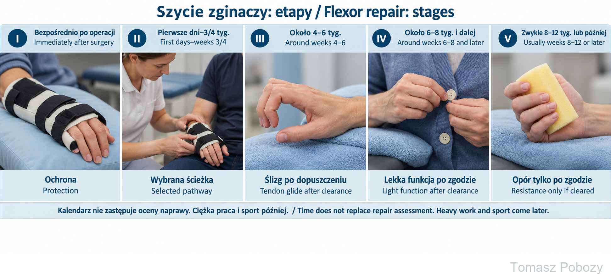
Stage I — immediate postoperative protection
Goal: protect the repair and establish a safe rehabilitation pathway.
Use the prescribed dorsal blocking orthosis and the hand position selected by the surgeon. Monitor swelling, sensation, and circulation. Move the shoulder and elbow if permitted. Finger motion is performed only within the selected pathway.
Avoid: forceful active flexion, strength testing, weight bearing through the hand, strong gripping, and lifting. Do not independently alter the orthosis or elastic tension.
Stage II — early motion or continued protection
Commonly from the first days to about weeks 3–4 — repair dependent
This period follows one of the pathways above. With Kleinert, extension is active only to the orthosis limit, without hyperextension; flexion is assisted by elastic bands running exclusively along the palmar side. Active finger flexion against elastic resistance is avoided. In Duran, flexion is passive. In early active motion, flexion is low-amplitude and tightly dosed.
Safety criteria: no sudden loss of active flexion, no escalating pain/swelling response, satisfactory sensation and perfusion, and reliable exercise technique.
Stage III — increasing tendon glide and active motion
Approximately weeks 4–6 — after healing review
Gradually increase active motion and differential tendon glide according to the selected programme. Do not force a full fist when tension is high, movement is poorly controlled, or the surgeon or therapist has not cleared that progression. Orthosis changes and removal should follow the treating plan.
Tendon gliding: finger-position sequences are introduced gradually, without pain and without forcing end range. Good passive motion with markedly poorer active motion can indicate adhesion-limited glide and requires assessment rather than an abrupt increase in exercise force.
Stage IV — reducing protection and developing function
Approximately weeks 6–8 onward — individualised
After healing review, gradually reduce orthosis use and increase light functional hand use. Develop fuller active motion, differential glide, and low-load tasks. Blocking exercises and more demanding glide work should be introduced only when compatible with the actual repair.
Stage V — strengthening and return to loading
Usually after weeks 8–12 or later — after explicit clearance
Begin resistance with low loads, for example gentle squeezing of a soft sponge or ball. Progress one variable at a time. Forceful grip, prolonged tool use, heavy manual work, and higher axial loads should come later than motion exercises.
Return to heavy work, fall-risk sport, and very forceful gripping is often around 12–16 weeks or later, but readiness should be based on motion quality, strength, tendon glide, and task demands rather than calendar time alone.
Warning signs
Warning signs and complications
Possible repair rupture: sudden loss of previously present active flexion, a snap/pop sensation, or abrupt functional deterioration. Stop exercises and seek urgent surgical review.
Other reasons for prompt assessment include increasing severe pain or swelling, new sensory disturbance, impaired finger perfusion, wound breakdown, or increasing redness. Later problems may include adhesion, contracture, and restricted tendon glide.
For a life-threatening emergency, call your local emergency number immediately — 112 in the European Union. On the boards, 112 refers to the EU.
Additional scope notes
HAND AND WRIST · FOR DOCTORS AND PHYSIOTHERAPISTS
Prepared by: Tomasz Poboży, MD, PhD — orthopaedic surgeon
Sources reviewed: 28 September 2026
Scope. This material is for adults after primary finger flexor tendon repair, particularly Zone II repair. It is not one schedule for every injury zone or pattern. Management changes with the number and type of repaired tendons, repair construct and strength, tissue quality, pulley status, associated nerve or vessel injury, fracture and additional procedures.
The operative record and the orthopaedic surgeon’s restrictions take priority over the timetable on this page.
Protect the repair first. Early motion is not a goal in itself. During the first weeks, management balances the risk of rupture or repair elongation against adhesion and stiffness. The motion strategy must match the actual strength of the repair and the patient’s ability to follow the programme safely.1–5, 7
May be appropriate when the repair needs additional protection, the injury is complex, or adherence is insufficient for a safe motion programme. Longer immobilisation, however, increases the risk of adhesion and stiffness.5, 7
A dorsal blocking orthosis protects the repair. Elastic traction runs on the palmar side and assists flexion; the patient actively extends only to the orthosis limit, without hyperextension. After relaxation, the elastic returns the fingers towards flexion.4, 6
Controlled passive motion aims to generate tendon glide while limiting active loading of the repair. Combined programmes are also used and should be matched to the repair construct.1, 5
Suitable only after an adequately stable repair, with reliable patient adherence and hand-therapy supervision. Meta-analysis found that a two-strand core repair was not sufficient for an active flexion-extension programme.2
References
References
- Starr HM, Snoddy M, Hammond KE, Seiler JG 3rd. Flexor tendon repair rehabilitation protocols: a systematic review. J Hand Surg Am. 2013;38(9):1712–1717.e14. doi:10.1016/j.jhsa.2013.06.025.
- Xu H, Huang X, Guo Z, et al. Outcome of Surgical Repair and Rehabilitation of Flexor Tendon Injuries in Zone II of the Hand: Systematic Review and Meta-Analysis. J Hand Surg Am. 2023;48(4):407.e1–407.e11. doi:10.1016/j.jhsa.2021.11.013.
- Mortada H, Alhithlool AW, Kattan ME, et al. Maximizing hand function following zone II flexor tendon repair: A systematic review and meta-analysis of rehabilitation strategies. J Hand Microsurg. 2024;16(5):100152. doi:10.1016/j.jham.2024.100152.
- Renberg M, Turesson C, Borén L, Nyman E, Farnebo S. Rehabilitation following flexor tendon injury in Zone 2: a randomized controlled study. J Hand Surg Eur Vol. 2023;48(8):783–791. doi:10.1177/17531934231166336.
- Chesney A, Chauhan A, Kattan A, Farrokhyar F, Thoma A. Systematic review of flexor tendon rehabilitation protocols in zone II of the hand. Plast Reconstr Surg. 2011;127(4):1583–1592. doi:10.1097/PRS.0b013e318208d28e.
- Werntz JR, Chesher SP, Breidenbach WC, Kleinert HE, Bissonnette MA. A new dynamic splint for postoperative treatment of flexor tendon injury. J Hand Surg Am. 1989;14(3):559–566. doi:10.1016/S0363-5023(89)80025-5.
- Kiwan E, Saad R, Najjar J, et al. Zone II Flexor Tendon Repair – Number of Strands, Technique, and Postoperative Rehabilitation: A Narrative Review. Orthop Rev (Pavia). 2026;18:161329. doi:10.52965/001c.161329.
References and links checked on 18 October 2026. Existence of a publication does not validate every dose or timetable. Institutional protocols and examples require individualisation.
Clinical commentary and terms of use
Commentary — one shared plan for surgeon and hand therapist
Rehabilitation should not be selected from the label “flexor tendon repair” alone. Temporary immobilisation, Kleinert/Duran, or early active motion depends primarily on the quality and construct of the repair and the patient’s ability to follow the programme safely. Contemporary reviews support controlled early motion after appropriately strong repairs but do not establish one universal protocol.1–5, 7