 Skip to content
FOR CLINICIANS AND PHYSIOTHERAPISTS
Total knee arthroplasty — rehabilitation protocol
From first standing to regaining knee control. Work on extension and flexion, muscle strength and safe walking is combined with progression tailored to healing and actual function.
Using and loading the limb
After stable primary TKA, WBAT with aids is often permitted. Cementless fixation alone does not prohibit full loading; follow the individual surgical plan.
Full guidance ↓Brace or immobilisation
A brace and CPM are not routine requirements after uncomplicated primary TKA. Additional repair may need separate protection.
Full guidance ↓Permitted movement
Restore extension and flexion gradually without forcing. Distinguish passive restriction from active extension lag; stage targets are not first-session goals.
Full guidance ↓Permitted movement, loading and removal of a brace are separate decisions. Additional repairs may require a more protective programme.
Rehabilitation at a glance
Progression follows healing and functional criteria, not dates alone.
Rehabilitation boards
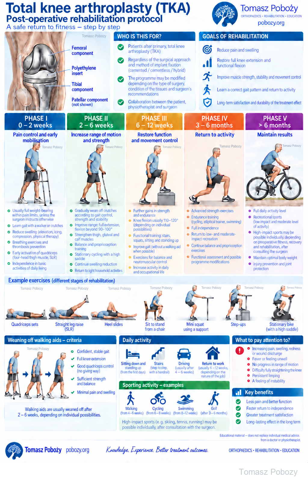
The patellar component is not shown in the implant diagram. The full protocol distinguishes early walking from later recreational walks.
Exercises — a closer look
Use the complete boards above together with the instructions for the chosen stage. An exercise shown for a later stage is not permitted early.
Open the relevant complete board and zoom in. The viewer retains all stages and captions.
Exercise instructions and progression are contained in the stage descriptions linked above.
Scope and protection
Protocol scope, surgical approach and weight-bearing
Adults following primary elective total knee arthroplasty, most often for osteoarthritis, with an uncomplicated recovery. The programme does not automatically apply to partial replacement, revision surgery, an acute fracture or additional ligament or extensor mechanism reconstruction.1
Before starting, establish permitted weight-bearing, movement restrictions, any additional repairs, wound condition and limb control after anaesthesia. Record preoperative range of motion, the condition of the other leg and the home environment.
Surgical approach and fixation
A standard medial parapatellar, midvastus or subvastus approach does not require a separate weekly schedule in this protocol. Tailor the programme to function and the documented restrictions. Closure of a standard arthrotomy is not equivalent to repair of a torn tendon. An additional extensor mechanism or ligament repair, or an osteotomy, may require separate protection.
With stable fixation, weight-bearing as tolerated with crutches or a walking frame is often permitted. A cementless implant does not automatically prohibit full weight-bearing. In a randomised trial of a particular TKA model, rehabilitation in both cemented and cementless groups included full weight-bearing from the day of surgery. Do not generalise this to unstable fixation, fracture or every implant model.4
Weight-bearing as tolerated allows loading of the operated leg to increase up to the level permitted by the orthopaedic surgeon. Absence of pain does not authorise exceeding a prescribed limit. Permission to walk does not authorise heavy resistance, sudden twisting or deep squats.
Pain, swelling and limb positioning
Pain management enables movement
The patient takes regular medicines according to the prescribed schedule and rescue medicines as needed. Selection accounts for contraindications, other illnesses and anticoagulant treatment. Do not delay baseline treatment until pain prevents exercise, or increase doses to force movement. Report inadequate analgesia to the team.5
Cooling may reduce symptoms and swelling. Protect the skin and monitor sensation and tissue response. Elevation and breaks in activity complement the programme. Positioning method and duration are individualised.1
Do not keep a pillow permanently under the knee, encouraging persistent flexion. This does not prohibit brief support during exercise or deliberate positioning to reduce swelling. During extension exercises, support the distal lower leg, protect the heel skin and do not force the knee down.
Walking and ankle pumps do not replace prescribed thromboprophylaxis. Do not independently change its duration or dose.2
Extension and flexion — how should poster goals be interpreted?
The numbers are indicative end-of-phase goals, rather than maximum permitted ranges or requirements for the first session. Uncomplicated TKA does not routinely require a six-week 90° flexion limit. Regain range without forcing and while protecting the healing wound.
The poster uses five phases: 0–2 weeks, 2–6 weeks, 6–12 weeks, 3–6 months and beyond 6 months. Phase II aims for full extension and gradual progression beyond 90–100° flexion; in phase III, functional flexion is often approximately 110–120°. This neither prohibits achieving a greater range earlier nor requires these values despite pain.
More detailed reference points can also help monitor the early weeks: approaching full extension and more than 70° flexion by the end of the first week, more than 90° around week four, and approximately 110° or more around week eight, if achievable without forcing. These are checkpoints, not separate treatment phases. Preoperative status and the actual trend of improvement matter.
Extension of 0° means neutral alignment, not hyperextension. Measure passive range, active range and active extension lag separately. If the knee can be straightened passively but the patient cannot maintain that position, regaining muscle control is also a goal. If passive extension is restricted too, work on mobility and assess the cause. Repeat measurements in a comparable position.3
Exercises and the daily home programme
The following exercises are examples, not a list to complete in full from day one. Following instruction, the patient may perform a selected set independently. The physiotherapist records the range, repetitions, frequency and rules for increasing difficulty.
Initial exercises
Quadriceps sets: support the leg, gently straighten the knee and tighten the front of the thigh. For example, hold for 5–10 seconds, then rest, without holding the breath. Heel slides: slide the foot towards the buttock, then return. The other leg or a strap may assist, without jerking.
Extension with lower-leg support: place support near the ankle, with no pillow under the bent knee; allow the relaxed leg to straighten gradually. Straight-leg raise: first tighten the quadriceps, then lift slightly; if extension is lost, make the task easier. Do not add an ankle weight for this test.
Functional exercises
Sit-to-stand: use a stable chair, higher and with armrests if needed. Low step: step up and lower slowly with a handrail. Mini-squat: use a worktop for support without letting the knee collapse inwards. Increase height, depth and resistance only with good control.1
A daily programme does not mean heavy training every day
Practise mobility, short walking distances and the activation exercises learned regularly at home. Resistance training has a separately selected frequency and recovery schedule. There is no need to delay every strengthening exercise until week six, but intensity must not exceed muscle control and tissue tolerance.6
In a trial comparing early higher- and lower-intensity training, both programmes improved function, with no demonstrated advantage of higher intensity in the main outcomes. Greater resistance is therefore not an end in itself. Adjust the dose according to pain, swelling, range and movement quality.6
After a session, also check the following day’s response. Clearly increased effusion, poorer gait or loss of gained range call for a lower dose and assessment. Mild, transient stretching discomfort does not necessarily mean damage; sharp or increasing pain is a reason to stop the task.
NMES, passive movement and adjunctive therapy
NMES — neuromuscular electrical stimulation: introduce early to supplement quadriceps activation. The 2026 guideline recommends at least daily use, at the highest tolerated intensity that produces an effective contraction. The physiotherapist assesses parameters, electrode placement and contraindications. Activation inhibition and active extension lag are particularly relevant.1
Passive, assisted and active movement are parts of the programme; they do not replace strengthening or gait training. Patellar and tissue mobilisation may support exercise, with attention to the wound. Do not routinely use a CPM machine or a brace solely to increase range after uncomplicated primary TKA. An exception following an additional repair or treatment of stiffness requires a separate prescription.1
Assessing progress and managing stiffness
Record pain at rest and during movement, swelling, wound condition, active and passive range, quadriceps activation, gait and need for assistance. Later, compare strength and task performance: chair rises, Timed Up and Go, walking and stairs. The other affected knee is not always a suitable reference for percentage symmetry.1, 6
Lack of progress is not an indication to force through resistance. If range fails to increase or decreases across successive reviews, investigate why. Practical reasons for earlier discussion with the surgeon include flexion still below approximately 90° at weeks 4–6 without an improving trend, or a persistent significant extension deficit. These do not automatically diagnose adhesions or indicate surgery.
Consider pain, effusion, wound problems, infection and mechanical causes, among others. When manipulation under anaesthesia is considered, timing matters — outcomes are often better within the first three months.7 This does not mean waiting three months to seek advice. The surgeon decides further treatment.
This protocol applies to uncomplicated primary TKA. Additional repairs, fractures or different fixation stability may alter the recommendations. Timings and ranges on the poster are indicative, rather than compulsory outcomes or movement limits. Recreational walking shown from weeks 4–6 does not mean waiting until then to start walking: initial mobilisation is described in phase I. Weight-bearing and return to sport must take account of the individual patient’s instructions.
Stages and progression criteria
Phase I · 0–2 weeks — pain control and early mobilisation
From first standing to basic independence
Rehabilitation starts on the day of surgery if the clinical condition allows, generally within 24 hours.1 Standing requires supervision, recovered limb control and tolerance of position changes. Absence of pain after a nerve block does not mean the patient is ready to walk independently.
Teach transfers, walking with aids and turning with small steps. Exercises include ankle pumps, quadriceps sets, heel slides and gentle extension. Perform straight-leg raises when the patient can maintain knee extension; if the knee drops into flexion, provide assistance and rebuild activation.
Before discharge: safe basic activities, stair competence if needed, an understandable home programme, controlled pain and an agreed route of contact with the team. A flexion measurement alone does not replace safety assessment.2
After returning home, continue short, regular movement sessions and walking distances with the necessary assistance. Work on extension and flexion while monitoring the wound, pain and swelling. End-of-phase goal: basic independence, safe walking with aids and gradual improvement in knee control.
Phase II · 2–6 weeks — mobility, strength and better walking
Improve active extension, chair rises and even weight transfer. Tailor permitted seated exercises, supported mini-squats and hip movements to limb control. Gradually introduce resistance and balance tasks without increasing effusion or losing technique.1
A stationary bicycle can provide gentle movement once mounting it and sitting on the saddle are safe. If flexion is insufficient, start with part-revolutions; do not force the pedal round. Smooth movement matters more than resistance initially.
Reduce walking assistance when the knee does not buckle under body weight and gait does not deteriorate after several passes. The indicative 2–6 weeks of crutch use shown on the poster is not a compulsory deadline for stopping them.
End-of-phase goal: work towards 0° extension, gradually improve flexion beyond 90–100°, reduce effusion and walk more confidently. Goals must not be achieved at the expense of healing.
Phase III · 6–12 weeks — restoring function and movement control
Increase exercise difficulty: seated knee extension with selected resistance, chair rises, low step-ups, mini-squats, and hip and calf exercises. Initially practise balance with stable support. Add load gradually without increasing effusion or losing technique.1
Also practise controlled lowering when stepping down. If the knee buckles suddenly, reduce step height or resistance, or use a handrail. Do not substitute excessive trunk lean for knee function.
Develop longer walks, cycling and safe single-leg tasks.1 Tailor exercises to planned work and living conditions. Greater fatigue should not cause limping, knee buckling or loss of control when turning. Pool exercises may be introduced once the wound has healed and clearance is given.
End-of-phase goal: maintained extension, functional flexion — often approximately 110–120° — improved strength, more efficient stairs and walking with the least necessary assistance. Twelve weeks elapsed does not automatically authorise running, jumping or full sports training.
Phase IV · 3–6 months — return to agreed activities
Continue strengthening, endurance and movement-control exercises. Gradually increase the difficulty of tasks needed for daily life and work. Prefer low-impact activities8: walking, cycling, swimming after wound healing and suitable recreation.
Return to a more demanding sport depends on previous skills, the course of treatment, function and a shared risk assessment. There is no single return month for everyone. Persistent limping, effusion or difficulty controlling the knee call for reassessment, rather than simply more exercises.8
End-of-phase goal: independence and tolerance of agreed activities without increasing symptoms or reinforcing compensatory movement patterns.
Phase V · beyond 6 months — maintaining gains
Maintain strength, endurance, balance and regular activity. Cycling on the poster is an example of recreation, not an exercise reserved until this phase.
Sports involving running, jumping or a high injury risk are not routinely recommended after TKA. Participation is assessed individually, according to preoperative function, the course of treatment and rehabilitation, and the type of activity. Individual clearance does not guarantee implant safety.8
Goal: lasting function that meets the patient’s needs. Rehabilitation does not automatically end on reaching a particular flexion measurement; continue developing strength and endurance.
Warning signs
When should crutches be stopped? Walking and daily activities
There is no single compulsory time to stop using crutches after TKA. After TKA, reduce assistance according to knee control, gait and balance. Progress from two crutches or a frame to one crutch on the opposite side, then to unaided walking if safe.
- Permitted weight-bearing allows support to be reduced.
- The knee does not buckle uncontrollably during stance; active extension is adequately controlled.
- Limping or trunk lean does not increase after several passes.
- The patient safely stands up, stops, turns and covers the required distance.
- Reducing support does not significantly worsen pain, swelling or function.
A correct straight-leg raise helps assessment but is not sufficient on its own to discontinue crutches. Support may be needed longer for extended walks or uneven ground. A negative Trendelenburg test does not replace assessment of knee control.
Stairs and daily activities
Initially, the more capable leg leads upstairs; downstairs, move the aid and operated leg first. Tailor the technique to both legs; later progress to alternating steps. Turn with small steps instead of twisting on a stationary foot.
Driving requires safe pedal control and emergency braking, with no effects from medicines that impair performance. The 4–6-week interval on the poster does not replace this assessment. Consider kneeling after wound healing, gradually and on a soft surface; it may be uncomfortable but is not automatically prohibited forever. Base the decision on the individual knee.
Warning signs
Urgent same-day assessment: new calf pain and swelling, fever or chills, wound discharge, increasing redness and pain.9, 10 Sudden inability to bear weight, loss of active extension after injury or new weakness or sensory changes also require stopping exercise and contacting the team.
Immediate emergency help: sudden shortness of breath, chest pain or fainting10; sudden pallor and coldness of the foot also require urgent help. In Poland, call 112. Do not wait for the next rehabilitation appointment.
Watch or listen
Is knee replacement always necessary? (Polish)
Tomasz Poboży discusses when knee replacement is considered. This material concerns treatment selection; use the protocol above for postoperative rehabilitation.
References
References
This is a synthesis of scientific guidelines and studies, rather than a literal translation of a single document. The schedule is indicative. Intervention principles incorporate the 2026 APTA update; older studies are used for specific topics, not presented as newer guidelines.
- Bove AM, Carroll LA, Cone S et al. Clinical practice guideline for physical therapist management of total knee arthroplasty: revision 2026. Physical Therapy. 2026;106(7):pzag058. doi:10.1093/ptj/pzag058.
- Wainwright TW, Gill M, McDonald DA et al. Consensus statement for perioperative care in total hip replacement and total knee replacement surgery: Enhanced Recovery After Surgery (ERAS®) Society recommendations. Acta Orthop. 2020;91(1):3–19. doi:10.1080/17453674.2019.1683790.
- Kittelson AJ, Elings J, Colborn K et al. Reference chart for knee flexion following total knee arthroplasty: a novel tool for monitoring postoperative recovery. BMC Musculoskeletal Disorders. 2020;21:482. doi:10.1186/s12891-020-03493-x.
- van der Lelij TJN, Marang-van de Mheen PJ, Kaptein BL et al. Continued Stabilization of a Cementless 3D-Printed Total Knee Arthroplasty: Five-Year Results of a Randomized Controlled Trial Using Radiostereometric Analysis. J Bone Joint Surg Am. 2023;105(21):1686–1694. doi:10.2106/JBJS.23.00221.
- Lavand’homme PM, Kehlet H, Rawal N, Joshi GP; PROSPECT Working Group. Pain management after total knee arthroplasty: PROcedure SPEcific Postoperative Pain ManagemenT recommendations. Eur J Anaesthesiol. 2022;39:743–757. doi:10.1097/EJA.0000000000001691.
- Bade MJ, Struessel T, Dayton M et al. Early High-Intensity Versus Low-Intensity Rehabilitation After Total Knee Arthroplasty: A Randomized Controlled Trial. Arthritis Care Res (Hoboken). 2017;69(9):1360–1368. doi:10.1002/acr.23139.
- Akhtar M, Razick D, Seibel A et al. Outcomes of Early Versus Delayed Manipulation Under Anesthesia for Stiffness Following Total Knee Arthroplasty: A Systematic Review and Meta-Analysis. J Arthroplasty. 2024;39(11):2872–2879. doi:10.1016/j.arth.2024.05.059.
- Lester D, Barber C, Sowers CB et al. Return to sport post-knee arthroplasty: an umbrella review for consensus guidelines. Bone Jt Open. 2022;3(3):245–251. doi:10.1302/2633-1462.33.BJO-2021-0187.R2.
- McNally M, Sousa R, Wouthuyzen-Bakker M et al. The EBJIS definition of periprosthetic joint infection: a practical guide for clinicians. Bone Joint J. 2021;103-B(1):18–25. doi:10.1302/0301-620X.103B1.BJJ-2020-1381.R1.
- Ishaaya E, Tapson VF. Advances in the diagnosis of acute pulmonary embolism. F1000Research. 2020;9:F1000 Faculty Rev-44. doi:10.12688/f1000research.21347.1.
Clinical commentary and terms of use
Commentary
After knee replacement, I would not judge rehabilitation by flexion degrees alone. Extension, effective quadriceps function and a leg the patient trusts during weight-bearing are also needed. Improved passive measurements are insufficient if the knee still buckles when walking.
I regard the ranges as a direction of work, not a result to achieve at any cost. A person with good preoperative mobility differs from someone with a longstanding contracture. Neither forcing a single deadline nor ignoring lack of progress for many weeks is useful.
The standard approach and fixation type do not replace information about the actual course of surgery. Additional repairs need protection. Conversely, I would not prolong unloading or prohibit exercises solely because of the label ‘cementless’ if fixation and recovery allow progression.
Cooperation in both directions
The physiotherapist helps develop the programme: examining function, selecting exercises and doses, educating the patient and assessing the response to exercise. The surgeon provides information about the procedure, stability and restrictions. Observations about progress, pain, effusion or extension difficulties should reach the surgeon, not only when a complication is suspected.
If individual variations cannot be agreed, follow the discharge instructions and the appropriate protocol. This still allows the physiotherapist to select methods independently within the agreed limits. A specific case, a specific patient, a specific surgeon.
Use of this protocol
© 2026 Tomasz Poboży - orthopaedic surgeon · pobozy.org. All rights reserved to the original text and poster.