 Skip to content
FOR CLINICIANS AND PHYSIOTHERAPISTS
ACL reconstruction — rehabilitation protocol
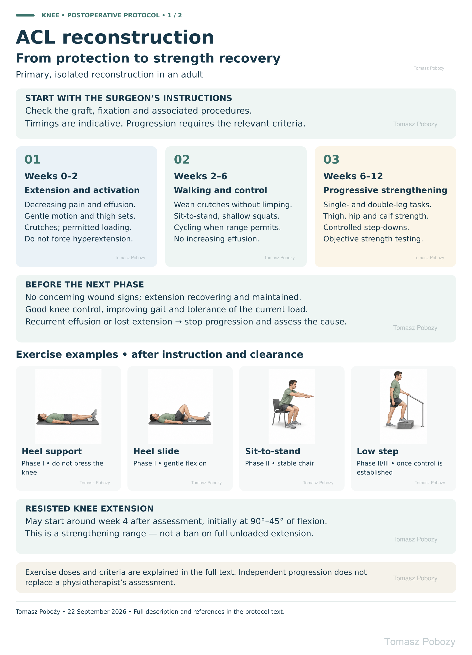
Rehabilitation after anterior cruciate ligament (ACL) reconstruction progresses from restoring knee extension and normal walking to rebuilding strength, running and a carefully planned return to activity. Two posters and a full written protocol describe management after primary, isolated ACL reconstruction in an adult. Progression requires assessment of knee function, the response to loading and the time needed for healing — the postoperative week alone is not sufficient.
Using and loading the limb
Isolated uncomplicated ACL reconstruction usually allows early progressive loading with crutches. Meniscal, cartilage or other procedures may change the limit.
Full guidance ↓Brace or immobilisation
Bracing is not identical for every patient. Settings and weaning follow discharge instructions, muscle control and additional repairs.
Full guidance ↓Permitted movement
Restore extension and permitted flexion early. The initial 90–45° resisted-extension range does not prohibit full extension without resistance.
Full guidance ↓Permitted movement, loading and removal of a brace are separate decisions. Additional repairs may require a more protective programme.
Rehabilitation at a glance
Progression follows healing and functional criteria, not dates alone.
Rehabilitation boards
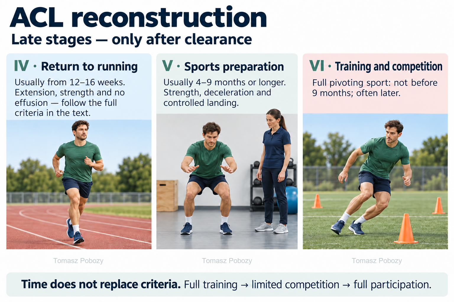
Stage IV: usually from 12–16 weeks after assessment. Stage V: usually 4–9 months or longer — strength, deceleration and landing. Stage VI: full pivoting sport not before 9 months, often later. Full training → limited competition → full participation. Time does not replace the criteria in the full protocol.
Exercises — a closer look · 3 boards
Use the complete boards above together with the instructions for the chosen stage. An exercise shown for a later stage is not permitted early.
Open the relevant complete board and zoom in. The viewer retains all stages and captions.
Exercise instructions and progression are contained in the stage descriptions linked above.
Stage IV: usually from 12–16 weeks after assessment. Stage V: usually 4–9 months or longer — strength, deceleration and landing. Stage VI: full pivoting sport not before 9 months, often later. Full training → limited competition → full participation. Time does not replace the criteria in the full protocol.
Scope and protection
Poradnik pacjenta: zerwane ACL — objawy i wybór leczenia (PL)
Scope, graft, weight-bearing and protection
What should be checked before starting?
Review the operation date and side, graft type and harvest site, fixation, associated procedures and prescribed restrictions on weight-bearing, motion and bracing. Assess the wound, effusion, extension, quadriceps activation, sensation and peripheral circulation. Establish the patient’s goal: daily function, manual work, running or pivoting sport. Missing operative information warrants contact with the orthopaedic surgeon, not selection of the fastest pathway.
Walking, crutches and bracing
After uncomplicated isolated reconstruction, movement and progressive weight-bearing as tolerated are usually introduced early, using two crutches and following postoperative instructions. Wean walking aids only when the patient controls the knee during stance, has no giving-way episodes and walks without a significant limp. A brace is not automatically required for the same duration in everyone: its settings, locking and discontinuation depend on the surgeon’s plan, muscle control and associated procedures. Do not rotate the trunk over a foot fixed on the ground; turn using several small steps. 1, 2
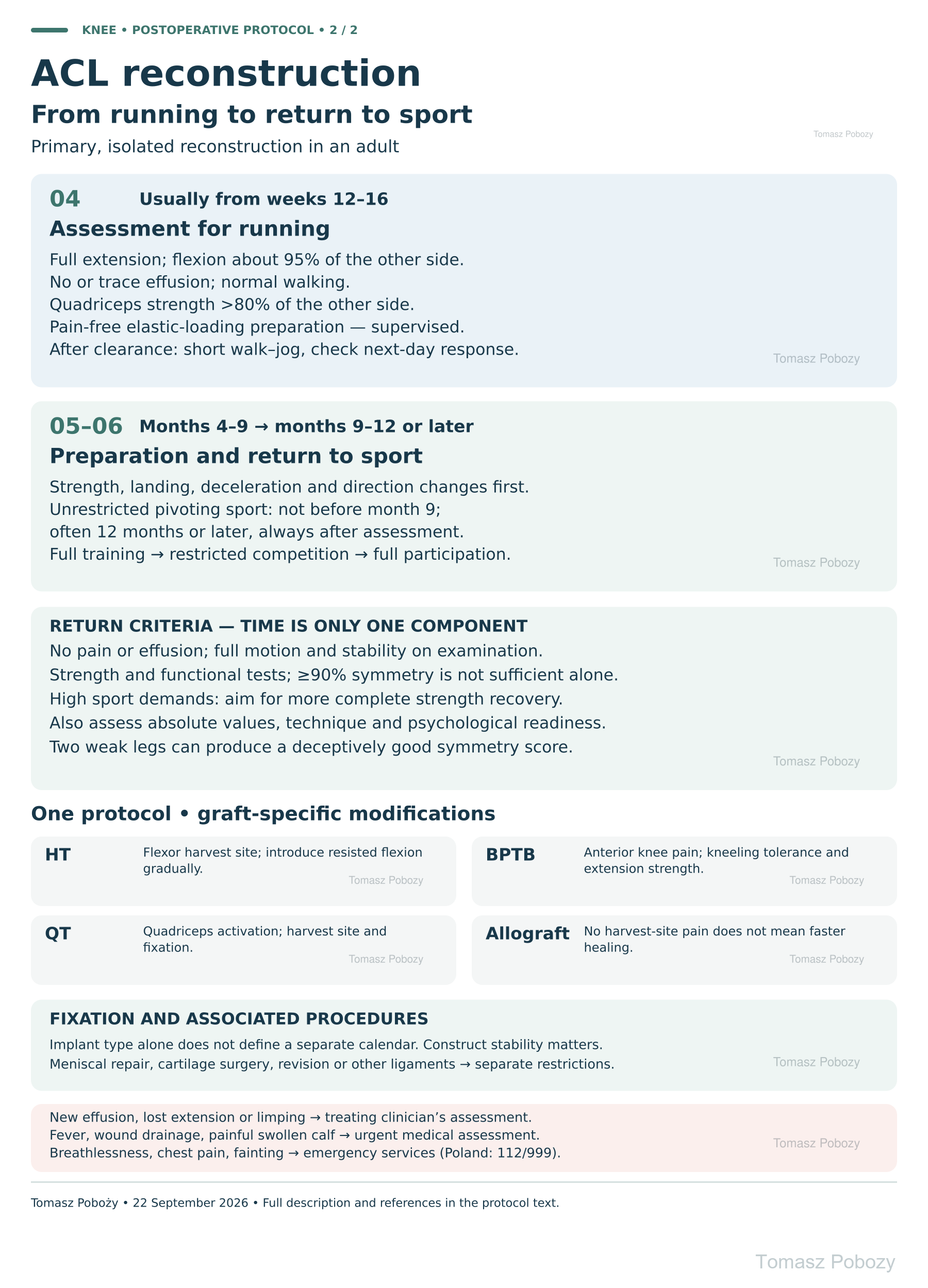
One base programme — graft-specific modifications
| Graft | Implications for rehabilitation |
|---|---|
| HT — hamstring tendon autograft | Assess harvest-site healing and knee-flexor strength. Introduce resisted knee flexion and high eccentric loads gradually, without provoking harvest-site pain. Do not apply one fixed-duration ban on hamstring exercises to every patient. |
| BPTB — bone–patellar tendon–bone autograft | Monitor anterior knee pain, kneeling tolerance and recovery of extension strength. Adjust squat depth and extensor-mechanism loading; do not abandon progressive strengthening. |
| QT — quadriceps tendon autograft, with or without a bone block | Pay particular attention to quadriceps activation and strength and the harvest-site response. Establish whether a bone block was harvested and how the graft was fixed; a large graft diameter does not automatically justify faster loading. |
| Allograft — donor tissue | Absence of harvest-site symptoms does not mean faster biological healing. Dynamic loading and return to sport generally warrant greater caution; the surgeon and rehabilitation team determine progression. |
These modifications reflect healing characteristics and harvest-site deficits; precise loading remains individual. 2, 3, 8
Fixation and associated procedures
The programme does not cover reconstruction with a synthetic ligament. Additional tape augmentation does not automatically permit faster rehabilitation — check the surgeon’s individual instructions.
Do not create a separate calendar solely from the name of a screw, cortical button, all-inside technique or number of bundles. Fixation matters: construct stability, bone quality and soft-tissue-to-bone or bone-block healing can alter permissible loading. However, there is no single sufficiently clinically validated timetable for every implant–graft combination. Restrictions documented by the surgeon take precedence. 3, 8
After meniscal repair, restrictions depend on the tear and repair type; root and radial tears often require particular protection. Do not automatically apply partial-meniscectomy precautions to a repaired meniscus. Cartilage surgery, osteotomy, additional ligament reconstruction or revision requires a plan covering all healing structures. An additional lateral procedure (LET/ALL) requires review of the instructions, but its name alone does not define a universal delay. 4
Exercises — technique and dosage
The doses below are author-proposed starting examples, not a compulsory routine for every patient. The physiotherapist selects exercises, range and resistance after examination and clearance for the relevant load. Brief mobility exercises can be distributed across several daily sessions; more demanding strength training is usually performed 2–3 times weekly with recovery time.
- Heel support and quadriceps set — phase I. Lie on your back with the heel on a low support and the knee unsupported. Let it straighten gently, without pressing with a hand or placing a weight on the knee. Then tighten the quadriceps for 5–10 seconds; for example, 8–12 repetitions. Do not force hyperextension.
- Heel slide — phase I. Slowly slide the heel towards the buttock on a smooth surface and back, without twisting the knee. For example, perform 8–12 gentle repetitions within the permitted range. A strap may assist the movement but must not be used for forceful pulling.
- Sit-to-stand — phase II, after weight-bearing clearance. Use a stable, initially higher chair. Place both feet on the floor, lean slightly forward and stand while keeping the knees controlled over the feet. Start with 1–2 sets of 8 repetitions; use support if needed. Avoid shifting all the work onto the uninjured leg.
- Low step — phase II/III, once control is established. Choose a step height appropriate to the patient; a rail provides safety. Place the whole foot on the step, rise slowly and control the descent. Initially use 1–2 sets of 6–10 repetitions, without the knee collapsing inward or pushing off with the other leg. Reduce height if technique deteriorates.
- Resisted knee extension — after assessment and clearance. Sit securely with the machine axis aligned with the knee. The initial range is described in section 2; an example dose is 2 sets of 10–15 repetitions with low resistance. Gradually change range and load while monitoring anterior knee and harvest-site pain. Later strengthening must become sufficiently challenging rather than remaining limited to light exercises.
- Knee-flexor, hip and calf strength — according to phase and graft. Select bridging, resisted knee flexion, hip exercises and calf raises to address deficits. After HT harvest, pay particular attention to harvest-site tolerance; after QT/BPTB, adapt extensor-mechanism work accordingly.
Response rule: mild exercise discomfort accepted by the clinician should not develop into sharp pain, increasing effusion, loss of extension or limping. If knee function is clearly worse the next day, reduce the dose and discuss an adjustment. For sudden deterioration, follow section 6.
Blood flow restriction (BFR) training is optional and should not be performed independently with an improvised strap. It may be considered as an adjunct under a trained practitioner after assessment of contraindications and risk. It does not replace progressive strength training. 1
Stages and progression criteria
The first 6 weeks — extension, control and walking
Phase I: usually weeks 0–2
Goals: decreasing pain and effusion, a healing wound, recovery of full extension, active quadriceps contraction and safe assisted walking. Check extension at every visit, comparing it with the other limb and the patient’s previous range. Do not force hyperextension with pressure.
Introduce ankle movements, gentle heel slides, quadriceps sets and gait practice. A straight-leg raise is appropriate if the knee stays straight; if an extension lag develops, reduce the difficulty and improve activation. Brief heel-supported positioning may help restore extension. Do not leave a pillow behind the knee for prolonged periods. Increase flexion gently, without forceful stretching or exceeding restrictions imposed by an associated procedure. 1, 2, 8
Cooling through a fabric layer and elevating the limb may ease symptoms; check skin and sensation and do not sleep with an ice pack in place. Follow discharge instructions for medication, dressings and thromboprophylaxis. Ankle exercises and walking do not replace prescribed prophylaxis. For quadriceps inhibition, the physiotherapist may add neuromuscular electrical stimulation (NMES). 1
Phase II: usually weeks 2–6
Goals: maintained extension, progressive flexion, good walking and stance control without increasing effusion. Progress closed-chain exercises: sit-to-stand from a higher chair, shallow squats, calf raises and a low step once adequate control is established. Introduce a stationary bicycle with low resistance when the wound and range permit, without forcing a full pedal revolution. Also train the hip, calf and trunk. 1, 2
Resisted knee extension is not prohibited throughout rehabilitation. In this protocol, controlled open-chain exercises may be considered from approximately week 4, initially between 90° and 45° of flexion, after clearance by the treating clinician. Adjust resistance and range progression to the graft, harvest site and symptoms. This range refers to a strengthening exercise, not a prohibition on recovering full unloaded extension. 1, 3
Progress when: the wound has no concerning signs, extension is full or approaching the individual target with any residual deficit explained, active knee control is good, walking is improving and the current exercise dose produces no significant deterioration. A persistent extension deficit warrants early assessment rather than waiting until the phase ends.
Strength recovery, running and return to sport
Phase III: usually weeks 6–12 — strength and single-leg control
Progressively load the quadriceps, knee flexors, hip muscles and calf. Combine open- and closed-chain exercises with concentric and eccentric work. Develop squatting, step-ups, step-downs and single-leg tasks without the knee collapsing inward or excessive trunk compensation. Adapt range and resistance to the joint and harvest-site response. Balance training alone does not replace strength recovery. 1, 2, 3
Measure strength objectively, for example with a belt-stabilised dynamometer or isokinetic testing. Record the method, position and result so that later measurements are comparable. Leg-press performance does not replace an isolated quadriceps strength measurement. 2
Phase IV: usually from approximately weeks 12–16 — assessment for running
Running does not start automatically at 3 months. Practical assessment includes full extension, flexion at approximately 95% of the other knee, no or only trace effusion, normal walking, quadriceps strength above 80% of the other side and pain-free tolerance of preparatory elastic-loading tasks. LSI is the operated-limb result divided by the non-operated-limb result × 100%. The clinician introduces hopping tests only after these loads have been cleared; patients should not jump to “test readiness” independently. If a force platform is available, assess eccentric work during jumping as well. These are supporting clinical criteria, not a test that guarantees graft safety. 1, 2
Start with short walk–jog intervals on a level surface, on separate days. An example starting dose after clearance is 1 minute of easy jogging / 2 minutes of walking, repeated 5 times. Increase tolerated duration before speed; do not increase volume, pace and terrain difficulty simultaneously. Effusion, limping or deterioration the following day warrants reducing the dose and reassessment.
Phase V: usually months 4–9 and beyond — sport preparation
After mastering earlier tasks, develop strength and power, double- and single-leg landing, acceleration, deceleration and planned, then reactive, changes of direction. Movement quality, load tolerance and sport demands determine order and difficulty. Easy running does not constitute clearance for competition, contact or sudden pivoting. Gradually reproduce sport situations and assess technique under fatigue as well. 2, 7, 9
Phase VI: usually months 9–12 or later — return to sport
For sports involving pivoting and abrupt deceleration, do not plan unrestricted return before month 9; many patients need 12 months or longer. Earlier return in young athletes was associated with a higher rate of subsequent injury; this is an observational finding, not a guarantee of safety once that time has passed. Clearance after an allograft or revision may be later. 2, 5
- A pain-free knee without effusion, full range of motion and stability confirmed by examination.
- Objective knee-extensor and flexor strength measurements and selected hop tests. At least 90% symmetry is a commonly used reference threshold, but not stand-alone clearance. For high-demand pivoting sports, aim for substantially more complete recovery; guidance for professional athletes proposes 100% isokinetic strength symmetry. 1, 2
- Also assess absolute values, preinjury results when available and sport-appropriate norms. Two weak legs can produce a deceptively good LSI. 6
- Effective deceleration, landing and direction changes without major compensations; tolerance of full training without a joint reaction the following day.
- Psychological readiness assessed by discussion and, as an adjunct, the ACL-RSI questionnaire; a shared decision by the patient, surgeon, physiotherapist and sports team. 7, 9
Progress through individual training → modified team training → full training → restricted competition → full participation. Continue strength training and secondary injury prevention after return. No single test eliminates the risk of another injury.
Daily activities and follow-up
Progress in everyday activities follows the permitted loading and motion for your procedure. Walking without aids requires a controlled gait; returning to driving requires safe vehicle control and no impairing medication. The detailed recommendations below explain the criteria and exceptions.
Daily activity, work and driving
Home, stairs and work
Remove slippery rugs and obstacles. Use stable support when sitting and standing; avoid sudden turns on the operated leg. Initially use stairs as taught by the physiotherapist, with a rail and crutches. Increase walking distance gradually without reinforcing a limp. Interrupt prolonged sitting with position changes and permitted movement.
Return to work depends on commuting, standing time, kneeling, lifting and slip risk. Desk work may be considered earlier than heavy manual work; remote work and gradually longer days can help. Demanding occupational tasks should be reproduced during rehabilitation before full clearance, just like sporting tasks. Do not prescribe one period of sick leave for everyone.
Driving
Driving is appropriate only when the patient can enter the vehicle, operate the pedals and perform rapid emergency braking safely, is not restricted by a brace and is not taking medication that impairs reactions. The operated side and transmission type matter. Several elapsed weeks alone do not establish readiness; when uncertain, seek the treating clinician’s assessment and check insurance conditions. 1
Water and recreation
Use a pool only once the wound has fully healed and the clinician has cleared it. Water exercise does not automatically permit forceful push-offs or a breaststroke kick. Stationary cycling precedes outdoor cycling, which introduces fall risk. Dancing, skiing and team sports require assessment for their specific loads, not simply completion of outpatient physiotherapy.
Warning signs
When to reduce loading and when to seek help
Stop progression and contact the treating clinician
Recurrent effusion, loss of previously recovered extension, new limping, harvest-site pain increasing across sessions or lack of progress require assessment of the cause and programme adjustment. Do not try to “walk off” a locked knee or aggressively stretch a painful extension deficit.
Urgent medical assessment
- Fever, worsening knee redness or warmth, wound drainage, severe increasing pain or marked general deterioration.
- New one-sided calf pain and swelling — suspected thrombosis requires urgent assessment; do not massage the calf.
- An injury with a pop, rapid swelling or instability; new locking.
- A cold or pale foot, new substantial sensory loss or foot weakness — seek immediate medical care.
Sudden breathlessness, chest pain, fainting or coughing blood: call emergency services — in Poland, 112/999; elsewhere, the local emergency number. Do not wait for a scheduled physiotherapy visit. This list does not cover every possible complication; rapid or unusual deterioration warrants medical assessment.
Additional scope notes
Start with the operative report and the surgeon’s instructions. The base programme applies to a stable, primary ACL reconstruction without an associated procedure requiring additional protection. Graft type and fixation modify loading decisions. Meniscal repair, cartilage surgery, multiligament reconstruction, revision surgery and skeletal immaturity require a separately agreed programme. This is not a protocol for primary ACL repair or a BEAR procedure. Timings are indicative; absence of pain does not establish graft strength.
References
References
- Kotsifaki R, Korakakis V, King E et al. Aspetar clinical practice guideline on rehabilitation after anterior cruciate ligament reconstruction. Br J Sports Med. 2023;57(9):500–514. doi:10.1136/bjsports-2022-106158.
- Brinlee AW, Dickenson SB, Hunter-Giordano A et al. ACL Reconstruction Rehabilitation: Clinical Data, Biologic Healing, and Criterion-Based Milestones to Inform a Return-to-Sport Guideline. Sports Health. 2022;14(5):770–779. doi:10.1177/19417381211056873.
- Solie B, Monson J, Larson C. Graft-Specific Surgical and Rehabilitation Considerations for Anterior Cruciate Ligament Reconstruction with the Quadriceps Tendon Autograft. Int J Sports Phys Ther. 2023;18(2):493–512. doi:10.26603/001c.73797.
- Pujol N, Giordano AO, Wong SE et al. The formal EU‐US Meniscus Rehabilitation 2024 Consensus: An ESSKA‐AOSSM‐AASPT initiative. Part I—Rehabilitation management after meniscus surgery (meniscectomy, repair and reconstruction). Knee Surg Sports Traumatol Arthrosc. 2025;33(8):3002–3013. doi:10.1002/ksa.12674.
- Beischer S, Gustavsson L, Senorski EH et al. Young Athletes Who Return to Sport Before 9 Months After Anterior Cruciate Ligament Reconstruction Have a Rate of New Injury 7 Times That of Those Who Delay Return. J Orthop Sports Phys Ther. 2020;50(2):83–90. doi:10.2519/jospt.2020.9071.
- Wellsandt E, Failla MJ, Snyder-Mackler L. Limb Symmetry Indexes Can Overestimate Knee Function After Anterior Cruciate Ligament Injury. J Orthop Sports Phys Ther. 2017;47(5):334–338. doi:10.2519/jospt.2017.7285.
- Meredith SJ, Rauer T, Chmielewski TL et al. Return to Sport After Anterior Cruciate Ligament Injury: Panther Symposium ACL Injury Return to Sport Consensus Group. Orthop J Sports Med. 2020;8(6):2325967120930829. doi:10.1177/2325967120930829.
- Wackerle AM, Sprague A, Joreitz R et al. Freddie Fu Panther Symposium Expert Group 2024: Rehabilitation and return to sport after anterior cruciate ligament reconstruction Part 1: Early and intermediate phases of rehabilitation. Knee Surg Sports Traumatol Arthrosc. 2026;34(2):520–530. doi:10.1002/ksa.70115.
- Wackerle AM, Joreitz R, Sprague A et al. Freddie Fu Panther Symposium Expert Group 2024: Rehabilitation and return to sport after anterior cruciate ligament reconstruction Part 2: Concepts and emerging technology in return to sport. Knee Surg Sports Traumatol Arthrosc. 2026;34(2):531–542. doi:10.1002/ksa.70110.
Clinical commentary and terms of use
Author’s commentary — why not one rigid timetable?
Reconstruction restores the ligament’s mechanical function, but does not complete treatment. Recovery of strength, movement control, exercise tolerance and confidence takes considerably longer than skin healing. Feeling well is not equivalent to completion of graft remodelling.
This publication uses one shared protocol with modifications. HT, BPTB, QT and allografts are not identical, so graft type is not omitted. However, separate, nearly repetitive pages organised by implant brand could imply a precision of timing that the evidence does not support. A clear record of restrictions arising from the actual operation is more important. A genuinely separate programme is needed when protecting another repaired structure, treating revision or multiligament reconstruction, or managing a skeletally immature patient. 3, 4, 8
The literature supports exercise-based rehabilitation and functional criteria, but does not establish one ideal dose or return-to-sport test for everyone. Some thresholds reflect expert consensus, while evidence on return-to-sport timing includes observational studies. The phase ranges, exercise selection and example doses here therefore form a practical synthesis for individualisation rather than reproduction of one study. 1, 2, 5, 7, 8, 9
Useful documentation includes the current phase, pain and effusion, extension and flexion, strength measurements, tolerated tasks, the next-day response and the condition for further progression. If considerable time has passed but criteria remain unmet, do not accelerate automatically — identify the cause.