 Skip to content
FOR CLINICIANS AND PHYSIOTHERAPISTS
Meniscal repair — rehabilitation protocol
From protecting the repair to restoring function. This protocol covers adults after primary repair of the medial or lateral meniscus. Tear pattern and the extent of repair determine early precautions; later progression depends on both healing time and knee function.
Using and loading the limb
Loading depends on tear and repair type. Early full loading may apply to stable vertical repairs; do not transfer it to radial or root repairs.
Full guidance ↓Brace or immobilisation
There is no single brace schedule for every repair. If prescribed, document walking lock, exercise range and weaning criteria.
Full guidance ↓Permitted movement
Unloaded flexion and squat depth have different limits. Follow repair-specific range for the first 6 weeks; avoid early twisting and deep loaded flexion.
Full guidance ↓Permitted movement, loading and removal of a brace are separate decisions. Additional repairs may require a more protective programme.
Rehabilitation at a glance
Progression follows healing and functional criteria, not dates alone.
Rehabilitation boards
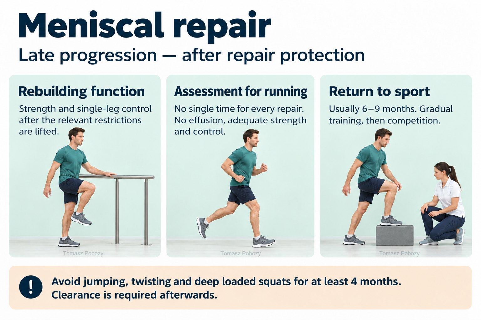
After the relevant restrictions are lifted: strength and single-leg control. There is no single time to start running after every repair; no effusion, adequate strength and control are required. Sport usually at 6–9 months: gradual training, then competition. Avoid jumping, twisting and deep loaded squats for at least 4 months; clearance is required afterwards.
Exercises — a closer look · 3 boards
Use the complete boards above together with the instructions for the chosen stage. An exercise shown for a later stage is not permitted early.
Open the relevant complete board and zoom in. The viewer retains all stages and captions.
Exercise instructions and progression are contained in the stage descriptions linked above.
After the relevant restrictions are lifted: strength and single-leg control. There is no single time to start running after every repair; no effusion, adequate strength and control are required. Sport usually at 6–9 months: gradual training, then competition. Avoid jumping, twisting and deep loaded squats for at least 4 months; clearance is required afterwards.
Scope and protection
Scope, weight-bearing and repair protection
What must be established before the first exercise?
Read the operative report for the side, meniscus and region, tear direction and extent, root involvement, repair method and stability, and associated procedures. Instructions should specify weight-bearing, range of motion, brace settings and follow-up. The label “all-inside repair” alone does not define the programme. Missing repair details require clarification rather than selection of the fastest pathway. 1
Longitudinal tear — the main pathway
After a stable vertical longitudinal repair, early full weight-bearing during walking may be permitted when prescribed by the orthopaedic surgeon. Full weight-bearing while walking does not permit squatting, pivoting on a planted foot or loading in deep flexion. Crutches remain necessary when there is a limp, poor knee control or inability to maintain the prescribed loading limit. 1
The consensus does not give one entirely consistent flexion limit: its table allows unrestricted motion for stable vertical repairs, while the text recommends limiting motion for 6 weeks after longitudinal repairs. These statements should not be averaged into an arbitrary limit. During the first 6 weeks, follow the surgeon’s prescribed range; clarify missing limits before progressing. Unloaded range of motion and permitted squat depth are separate instructions. 1
Repairs requiring additional protection
| Repair type | Early loading and movement |
|---|---|
| Complete radial or oblique tear | Consensus: no weight-bearing for 4–6 weeks and motion of 0–90° for 4–6 weeks. The surgeon determines the specific duration. |
| Horizontal tear in a young athlete | Consensus: partial or no weight-bearing and motion of 0–90° for the first 4 weeks. Do not extrapolate automatically to extensive degenerative repairs. |
| Complex / extensive vertical tear | Written restrictions are required, commonly protecting motion and/or loading for 4–6 weeks. The consensus permits full weight-bearing in some complex vertical repairs; not every complex repair is equivalent. |
| Meniscal root | Separate programme: the consensus recommends no weight-bearing for 6 weeks. For 0–90° motion, the table specifies 4 weeks and the discussion 6 weeks. Do not accelerate without clarification in the surgeon’s instructions. |
| Ramp lesion / repair with ACL reconstruction | The ligament procedure also determines the plan. Meniscal restrictions may slow early ACL progression, while ACL reconstruction may delay return to sport. |
These distinctions come from the consensus and do not authorise independent changes to discharge instructions. Medial versus lateral location alone does not determine a different timetable; the repaired tear pattern matters more. 1
Bracing
There is no single established bracing regimen for all meniscal repairs. If a brace is prescribed, clarify its settings for walking, exercise and rest, and the criteria for unlocking and discontinuing it. Unlocking a brace does not itself permit greater loading. 1
Exercises — technique and dose selection
The following instructions explain exercise technique; they are not a mandatory set for every patient. Research has not established one optimal set-and-repetition prescription for all repairs. After assessment, the physiotherapist records range, resistance, repetitions and frequency; the dose should not cause increasing effusion or deteriorating function.
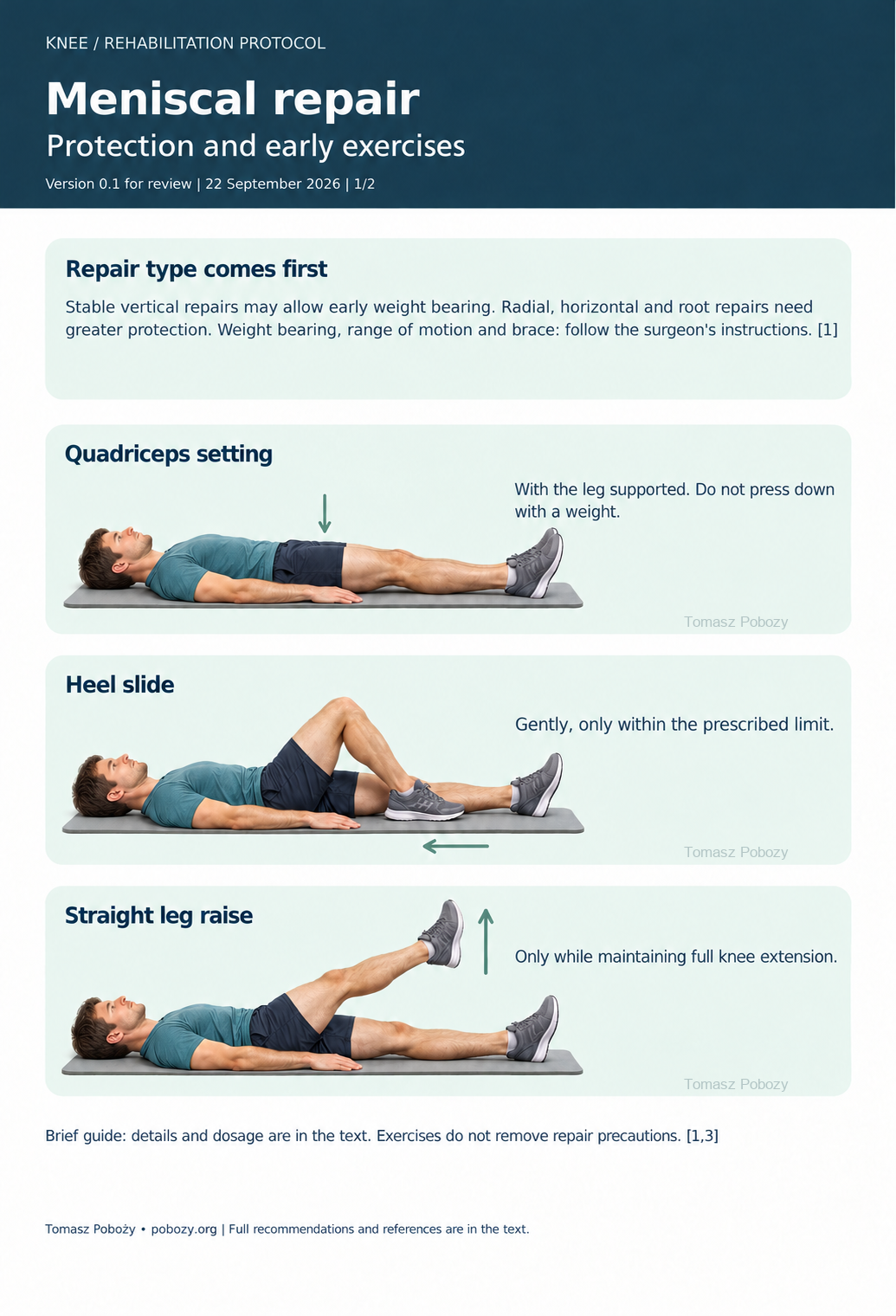
A. Quadriceps set
Lie with the leg supported and tighten the front of the thigh without lifting the heel. Breathe normally and relax before repeating. Do not press the knee down with an external weight. 3
B. Assisted heel slide
Slide the heel on a smooth surface to gently bend the knee, then return towards extension. A strap or the other leg may assist, without forcing flexion. Stop at the prescribed limit and avoid rotating the foot. 1
C. Straight-leg raise
Activate the quadriceps first. Lift only while maintaining knee extension, then lower slowly. Stop if the knee sags into flexion and return to activation exercises. 3
D. Supported mini-squat — only after clearance
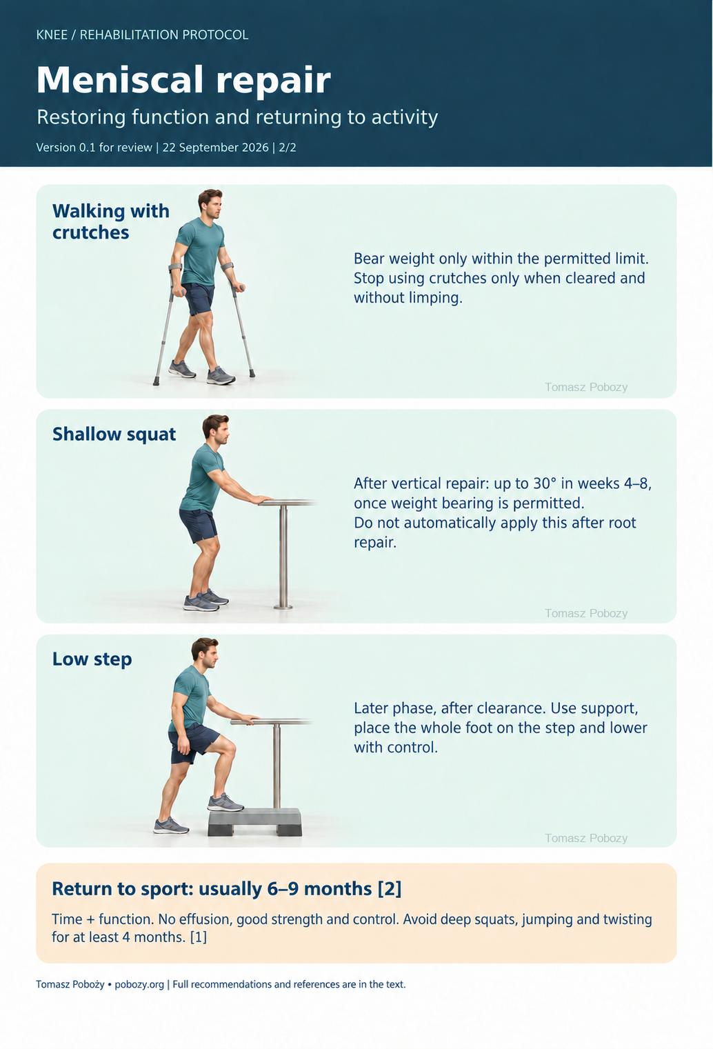
Stand at a stable support with both feet on the ground. Move the hips back and bend the knees only to the permitted depth. Keep the knees aligned with the feet without twisting the trunk. Return smoothly to standing. Section 3 gives the angular limits for longitudinal repairs. 1
E. Controlled low step-up — a later-phase exercise
After clearance for this load, place the whole foot on a low step while holding a rail. Step up without pushing off with the other leg and lower slowly. Step height must fit the permitted knee flexion; reduce difficulty if pain or inward knee collapse occurs. 1
F. Stationary cycling and complementary training
Introduce cycling when the permitted range allows smooth pedalling without forced flexion. Begin with low resistance. Resisted knee-flexor exercise requires separate prescription, especially after posterior horn, radial or root repair. Pain-free cycling is not a test of healing. 1,3
Stages and progression criteria
Protection phase — surgery until precautions are lifted
The first days
Assess the wound, pain, effusion, extension, peripheral sensation and circulation, and quadriceps activation. Teach safe transfers and crutch-assisted walking within prescribed weight-bearing limits. Follow discharge instructions for medication, dressings and thromboprophylaxis. Alternate short bouts of permitted movement with rest; do not increase walking at the expense of a persistent limp.
Early exercises include gentle ankle movement, quadriceps sets and controlled knee flexion and extension within the permitted range. Avoid prolonged support directly behind the knee. Recover extension without forceful pressure or forced hyperextension. Introduce a straight-leg raise when the patient can maintain extension without a lag. 1,3
The remaining protection period
Reduced pain alone does not permit more weight-bearing. Continue motion within prescribed limits, quadriceps activation, and hip and trunk exercises that do not transmit prohibited loads to the operated knee. Supervised NMES may be considered for persistent muscle inhibition. 1
Once full weight-bearing is permitted, wean crutches according to gait quality and knee control. Effusion, joint-line pain or worsening gait after an activity increase calls for reduced loading and reassessment. Range progression must respect repair protection. 1
Criteria for entering the restorative phase
- The prescribed protection period has ended, with clearance to increase loading and range of motion.
- Full or nearly full passive range achieved without breaching earlier restrictions.
- No effusion and good quadriceps control.
- Safe walking consistent with current instructions.
The consensus identifies motion, absence of effusion and neuromuscular control as progression criteria. Do not force flexion merely to pass a milestone before protection ends. 1
Strength, running and return to sport
Progressive strengthening
Once the relevant restrictions are lifted, develop thigh, hip and calf strength, balance and control during stance. Progress from bilateral to single-leg tasks and from lower to greater resistance. Use both supported weight-bearing tasks and unloaded exercise as appropriate. Monitor the joint response after training and the following day. 1
For longitudinal repairs, the consensus proposes mini-squats up to 30° during weeks 4–8, up to 45° during weeks 8–12, and gradually to 60–90° during weeks 13–16. These are expert recommendations, conditional on loading clearance and good control. Do not automatically apply them after radial or root repair. Avoid deep loaded squatting, jumping and rotational knee activities for at least 4 months. 1
Entering preparation for activity
Assess full active range, objectively measured strength and dynamic single-leg knee control. The consensus gives approximately 80% of the other limb’s strength as a supporting threshold for entering the next phase. This is not discharge from rehabilitation or standalone clearance to run, jump or compete. Consider movement quality and absolute performance too; the other limb may also be weak. 1
Running
There is no single well-established starting week for running after every repair. Consider repair type and healing time, absence of effusion or mechanical symptoms, full motion and adequate strength and control. After clearance, start with short walking and jogging intervals on a level surface. Increase tolerated volume before speed and difficulty. Do not start with sprinting or cutting. 1,2
Full sport
The consensus generally recommends return to sport 6–9 months after meniscal repair, with assessment of function, strength, effusion, sport tasks and psychological readiness. The minimum 4 months of rehabilitation described for some vertical repairs is not automatic clearance for unrestricted competition. ACL reconstruction or other associated surgery may delay return further. 1,2
- Progress through preparatory tasks, individual practice, fuller sport-specific training and then competition.
- Measure strength with a reproducible method. Introduce hop testing only after impact loading has been cleared.
- Check the response to full training. Recurrent effusion or locking prevents appropriate progression.
No single test guarantees meniscal healing or freedom from another injury. 1,2
Daily activities and follow-up
Progress in everyday activities follows the permitted loading and motion for your procedure. Walking without aids requires a controlled gait; returning to driving requires safe vehicle control and no impairing medication. The detailed recommendations below explain the criteria and exceptions.
Daily activities and work
Remove obstacles and slippery rugs at home. Rise from a stable seat using armrests or other secure support. Turn with several small steps instead of twisting over a planted foot. If non-weight-bearing is prescribed, do not load the operated leg during transfers.
Use the stair technique taught by the physiotherapist, with a rail and appropriate crutch assistance. Managing essential stairs does not mean step training is cleared. Kneeling, deep squatting and twisting work require later clearance consistent with repair protection. 1
Return to work depends on demands: sitting with opportunities to change position differs from prolonged standing, lifting or kneeling. Arrange modified duties and gradual increases in activity where needed. Rehearse demanding occupational tasks during rehabilitation before unrestricted return.
Driving requires reliable, rapid pedal operation and emergency braking. Do not drive when limited by a brace, weight-bearing restriction, pain or medication that impairs reactions. The operated side and transmission matter; elapsed time alone does not establish driving fitness.
Pool activity may be considered after complete wound healing and clearance. Immersion does not remove repair restrictions: push-offs, twisting and breaststroke kicks are not automatically permitted. Increase activity without causing recurrent effusion.
Warning signs
When to stop progression and seek help
Reassessment by the treating team
Recurrent effusion, joint-line pain, lost motion, limping, painful catching or locking require assessment. Do not try to walk through a locked knee. If rehabilitation milestones remain unattainable, establish the reason before increasing the load. 1,2
Urgent medical assessment
- Fever, wound discharge, increasing redness, severe pain or rapid deterioration may indicate infection or another complication.
- New unilateral calf swelling and pain require urgent assessment for thrombosis; do not massage the calf.
- A cold or pale foot, marked new sensory loss or weakness of foot movement require immediate assessment.
- Sudden breathlessness, chest pain, fainting or coughing blood require emergency help — call 112/999 in Poland or the local emergency number.
Ankle movements and permitted walking do not replace prescribed thromboprophylaxis. This is not an exhaustive list of complications; rapid or unusual deterioration requires medical assessment.
Additional scope notes
The main pathway concerns an isolated longitudinal tear repair. Radial, horizontal, complex and root repairs require the additional precautions described below. Root reattachment, meniscal transplantation, revision surgery, osteotomy and cartilage repair are not covered by one common timetable. With concomitant ACL reconstruction, the requirements of both procedures must be met.
References
References
- Pujol N, Giordano AO, Wong SE, et al. The formal EU-US Meniscus Rehabilitation 2024 Consensus. An ESSKA-AOSSM-AASPT Initiative. Part I – Rehabilitation Management After Meniscus Surgery (Meniscectomy, Repair, Reconstruction). Int J Sports Phys Ther. 2025;20(6):918–930. doi:10.26603/001c.137095.
- Prill R, Ma CB, Wong SE, et al. The formal EU-US Meniscus Rehabilitation 2024 Consensus: An ESSKA-AOSSM-AASPT initiative. Part II—Prevention, non-operative treatment and return to sport. Knee Surg Sports Traumatol Arthrosc. 2025;33(8):3014–3024. doi:10.1002/ksa.12689.
- Monson JK, Tollefson LV, LaPrade CM, LaPrade RF. Current Rehabilitation Principles Following Meniscus Repairs. Curr Rev Musculoskelet Med. 2025;18:331–343. doi:10.1007/s12178-025-09967-6.
- Bouchard MD, Macciacchera M, Gilbert J, Lameire DL, Abouali J. Postoperative Rehabilitation Protocol After Isolated Meniscal Repair: A Systematic Review. Orthop J Sports Med. 2025;13(7):23259671251357513. doi:10.1177/23259671251357513.
- Albishi W, Al Yami I, Alyami A, Aldosari OA, AlJasser S. Rehabilitation Strategies Following Isolated Meniscal Repair: A Systematic Review of Protocols and Outcomes. J Clin Med. 2026;15(4):1616. doi:10.3390/jcm15041616.
Clinical commentary and terms of use
Commentary
Evidence and remaining uncertainty
The framework follows the 2024 ESSKA–AOSSM–AASPT consensus, published in 2025. Most recommendations after repair rely on limited evidence and expert opinion. No complete programme has been shown superior to all alternatives. Read time frames and functional criteria together. 1,2
The 2025 review by Bouchard and colleagues mainly included longitudinal or vertical tears. Heterogeneity and the predominance of non-randomised studies prevent a conclusion that early full weight-bearing is equally safe for every repair. These findings should not be extrapolated directly to root repairs. 4
The 2026 review by Albishi and colleagues also reports heterogeneous programmes. Its search covered publications through May 2024. 5
Record repair type, current precautions, pain and effusion, motion, strength measurements, tolerated tasks and the next progression criterion. A separate surgeon’s commentary should describe the actual case; a general protocol cannot replace the operative report.