 Skip to content
FOR CLINICIANS AND PHYSIOTHERAPISTS
De Quervain release surgery — First dorsal compartment release — rehabilitation protocol
Care after isolated open first dorsal compartment release in an adult. Protect healing tissues, restore gentle thumb and wrist movement and gradually return to gripping and work.
Using and loading the limb
Initially avoid forceful grip, wringing and lifting with a widely abducted thumb. Thumb resistance and greater loads follow assessment.
Full guidance ↓Brace or immobilisation
Both movement without splinting and short thumb protection are described after isolated release. The surgeon selects type, duration and removal instructions.
Full guidance ↓Permitted movement
Use permitted gentle thumb and wrist motion without forcing end range. Do not use the Finkelstein test as an intensive stretching exercise.
Full guidance ↓Permitted movement, loading and removal of a brace are separate decisions. Additional repairs may require a more protective programme.
Rehabilitation at a glance
Progression follows healing and functional criteria, not dates alone.
Rehabilitation boards
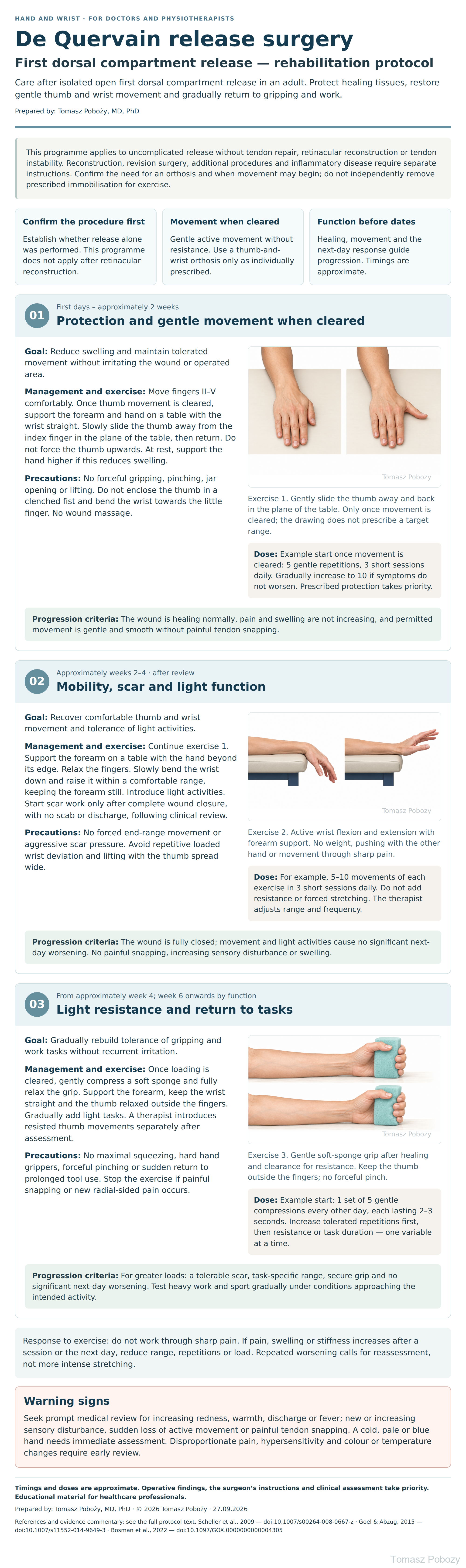
Exercises — a closer look
Use the complete boards above together with the instructions for the chosen stage. An exercise shown for a later stage is not permitted early.
Open the relevant complete board and zoom in. The viewer retains all stages and captions.
4. Exercise technique and dose selection
Thumb: keep the hand supported. Slide the thumb sideways on the table, then return to a relaxed position. Do not press it against the index finger. The range may be small. Avoid elevating the shoulder or rotating the forearm to create the appearance of more movement.
Wrist: the table edge supports the forearm without pressing on the operated area. Move the hand down and up without a weight. Keep the fingers relaxed. Do not swing the hand using momentum.
Grip: use an easily compressible sponge. Do not tuck the thumb under the fingers. Alternate a brief, gentle squeeze with complete relaxation. Grip exercise does not replace individually selected thumb strengthening.
Response to exercise: do not work through sharp pain. If pain, swelling or stiffness increases after a session or the next day, reduce range, repetitions or load. Repeated worsening calls for reassessment, not more intense stretching.
The repetition counts and frequency are proposed cautious starting doses for clinical review, not a programme validated in a trial after this operation. Stretching with the thumb enclosed in a fist and the wrist deviated towards the ulna is not a routine early exercise.
Scope and protection
1. Scope, initial assessment and orthosis
This programme applies to uncomplicated release without tendon repair, retinacular reconstruction or tendon instability. Reconstruction, revision surgery, additional procedures and inflammatory disease require separate instructions. Confirm the need for an orthosis and when movement may begin; do not independently remove prescribed immobilisation for exercise.
The release addresses the compartment containing the abductor pollicis longus (APL) and extensor pollicis brevis (EPB) tendons. Confirm the side, operative report, any septa and additional procedures. Record surgical restrictions and work demands. Assess the wound, swelling, pain, sensation, circulation and thumb and wrist movement; assess strength only once loading is permitted.
A published series used immediate movement without splinting after isolated release.1 A rehabilitation review instead described a thumb-spica orthosis and initiation of movement therapy at 2 weeks.2 Neither approach should be imposed on everyone. If an orthosis is prescribed, confirm wearing time, permitted breaks and movement. Obtain missing instructions from the orthopaedic surgeon before thumb and wrist exercises.
Stages and progression criteria
2. Phase I — protection and permitted movement
First days – approximately 2 weeks: Protection and gentle movement when cleared
Goal: Reduce swelling and maintain tolerated movement without irritating the wound or operated area.
Management and exercise: Move fingers II–V comfortably. Once thumb movement is cleared, support the forearm and hand on a table with the wrist straight. Slowly slide the thumb away from the index finger in the plane of the table, then return. Do not force the thumb upwards. At rest, support the hand higher if this reduces swelling.
Dose: Example start once movement is cleared: 5 gentle repetitions, 3 short sessions daily. Gradually increase to 10 if symptoms do not worsen. Prescribed protection takes priority.
Precautions: No forceful gripping, pinching, jar opening or lifting. Do not enclose the thumb in a clenched fist and bend the wrist towards the little finger. No wound massage.
Progression criteria: The wound is healing normally, pain and swelling are not increasing, and permitted movement is gentle and smooth without painful tendon snapping.
3. Phases II–III — movement, light resistance and function
Approximately weeks 2–4 · after review: Mobility, scar and light function
Goal: Recover comfortable thumb and wrist movement and tolerance of light activities.
Management and exercise: Continue exercise 1. Support the forearm on a table with the hand beyond its edge. Relax the fingers. Slowly bend the wrist down and raise it within a comfortable range, keeping the forearm still. Introduce light activities. Start scar work only after complete wound closure, with no scab or discharge, following clinical review.
Dose: For example, 5–10 movements of each exercise in 3 short sessions daily. Do not add resistance or forced stretching. The therapist adjusts range and frequency.
Precautions: No forced end-range movement or aggressive scar pressure. Avoid repetitive loaded wrist deviation and lifting with the thumb spread wide.
Progression criteria: The wound is fully closed; movement and light activities cause no significant next-day worsening. No painful snapping, increasing sensory disturbance or swelling.
From approximately week 4; week 6 onwards by function: Light resistance and return to tasks
Goal: Gradually rebuild tolerance of gripping and work tasks without recurrent irritation.
Management and exercise: Once loading is cleared, gently compress a soft sponge and fully relax the grip. Support the forearm, keep the wrist straight and the thumb relaxed outside the fingers. Gradually add light tasks. A therapist introduces resisted thumb movements separately after assessment.
Dose: Example start: 1 set of 5 gentle compressions every other day, each lasting 2–3 seconds. Increase tolerated repetitions first, then resistance or task duration — one variable at a time.
Precautions: No maximal squeezing, hard hand grippers, forceful pinching or sudden return to prolonged tool use. Stop the exercise if painful snapping or new radial-sided pain occurs.
Progression criteria: For greater loads: a tolerable scar, task-specific range, secure grip and no significant next-day worsening. Test heavy work and sport gradually under conditions approaching the intended activity.
Daily activities and follow-up
Daily activities follow the permitted joint motion, lifting, grip and arm support for your procedure. Ending immobilisation is not clearance for lifting or driving. The detailed guidance below explains the criteria and exceptions.
5. Daily activities, work, sport and driving
Start with short, light activities with the wrist close to neutral. Limit prolonged thumb typing, wringing, opening tight jars and carrying heavy bags. When caring for a child, share the load between both arms and forearms; do not lift using widely spread thumbs alone.
Computer work, playing an instrument, racket sport and heavy manual work have different demands. First increase the duration of short, tolerated tasks, then force and repetition. Reaching 6 weeks alone does not establish readiness for every load.
Driving requires a secure grip and ability to perform an emergency manoeuvre without significant pain or medication-related impairment. An orthosis that restricts vehicle control prevents a safe return. Consider the treating clinician’s instructions and vehicle demands.
Warning signs
6. Pain, warning signs and lack of progress
Seek prompt medical review for increasing redness, warmth, discharge or fever; new or increasing sensory disturbance, sudden loss of active movement or painful tendon snapping. A cold, pale or blue hand needs immediate assessment. Disproportionate pain, hypersensitivity and colour or temperature changes require early review.
Do not intensify exercise to overcome painful snapping. Persistent pain may require assessment of completeness of release, the scar, superficial radial nerve, tendon stability and coexisting problems such as thumb-base joint disease.3 Early review is also needed if complex regional pain syndrome is suspected.
Monitor the trend in symptoms and function. If improvement is absent across follow-up visits, revisit the clinical assessment and operative report. Take medicines as individually prescribed; do not increase doses to force training.
For a life-threatening emergency, call your local emergency number immediately — 112 in the European Union. On the boards, 112 refers to the EU.
Additional scope notes
References
8. References
The publications support management principles and outcome assessment. They do not establish one compulsory weekly schedule or repetition count.
- Scheller A, Schuh R, Hönle W, Schuh A. Long-term results of surgical release of de Quervain’s stenosing tenosynovitis. Int Orthop. 2009;33(5):1301–1303. doi:10.1007/s00264-008-0667-z.
- Goel R, Abzug JM. de Quervain’s tenosynovitis: a review of the rehabilitative options. Hand (N Y). 2015;10(1):1–5. doi:10.1007/s11552-014-9649-3.
- Bosman R, Duraku LS, van der Oest MJW, et al. Surgical Treatment Outcome of de Quervain’s Disease: A Systematic Review and Meta-analysis. Plast Reconstr Surg Glob Open. 2022;10(5):e4305. doi:10.1097/GOX.0000000000004305.
References and links checked on 18 October 2026. Existence of a publication does not validate every dose or timetable. Institutional protocols and examples require individualisation.
Clinical commentary and terms of use
7. Commentary — evidence and individualisation
Individual case, individual patient, individual surgeon. Available publications do not establish one best postoperative programme. The Scheller series describes early movement after a particular release technique, not a universal recommendation for reconstruction.1
Goel and Abzug describe their own programme: immediate postoperative splinting, movement therapy at 2 weeks and gentle strengthening at 4 weeks. This is a review and an account of the authors’ practice, not a comparative exercise-dosing trial.2 The time windows here are reference points, and the proposed doses require adjustment.
A systematic review supports generally favourable surgical outcomes but identifies complications and limitations in the quality of many studies; it does not validate a specific repetition count.3 The surgeon communicates procedure details and protection requirements; the hand therapist or physiotherapist assesses function, teaches technique and grades loading. A painful scar, restricted movement and demanding work warrant closer supervision.