 Skip to content
FOR CLINICIANS AND PHYSIOTHERAPISTS
Distal biceps tendon repair — rehabilitation protocol
Protect the reattachment at the radial tuberosity, restore movement, then rebuild elbow flexion and forearm supination strength. For doctors and physiotherapists.
Using and loading the limb
Initially no lifting or body-weight support. Active motion clearance does not permit biceps resistance; follow one fixation-specific programme.
Full guidance ↓Brace or immobilisation
Brace type, extension block and duration depend on repair tension and the surgeon. Do not average different published protocols.
Full guidance ↓Permitted movement
Protected flexion and supination may be passive, assisted or active according to the selected pathway. Do not force extension or independently test strength.
Full guidance ↓Permitted movement, loading and removal of a brace are separate decisions. Additional repairs may require a more protective programme.
Rehabilitation at a glance
Progression follows healing and functional criteria, not dates alone.
Rehabilitation boards
BOARD 01
From protection to loading
Weeks organise the stages but do not automatically authorise progression. Active movement and resistance exercise require separate assessment; absence of pain alone does not confirm healing.
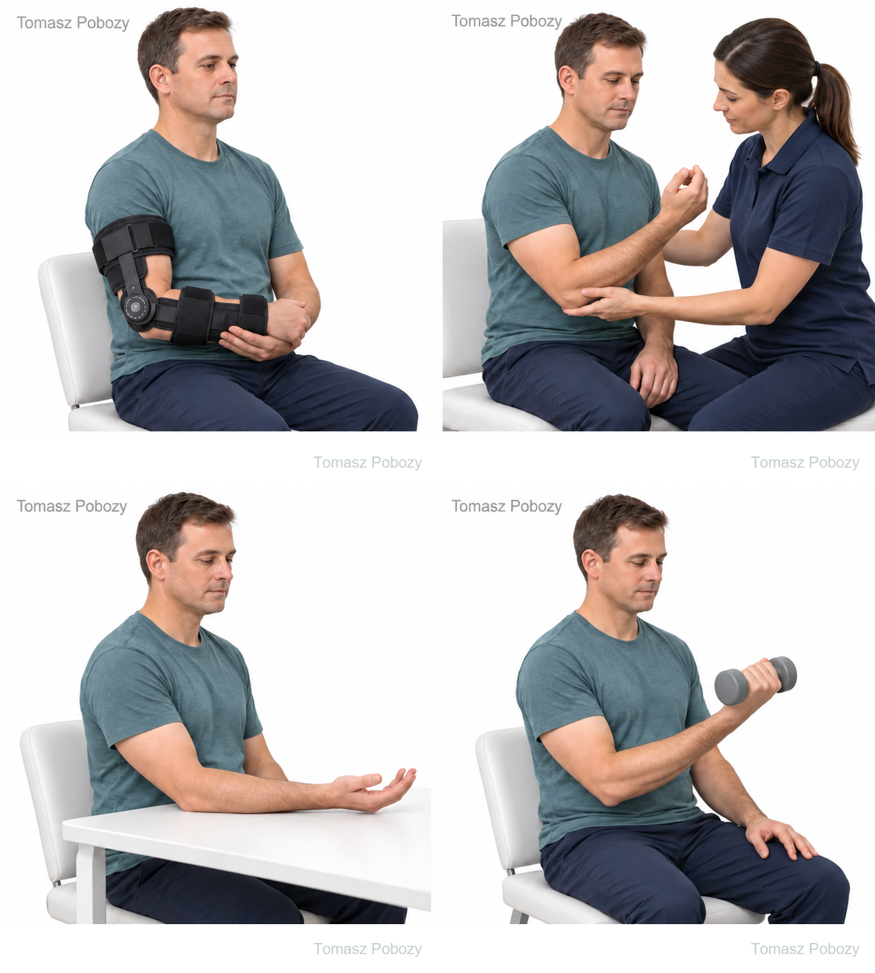
02 · Weeks 2–6
Protected movement
Gradually increase the permitted range without resistance. Use passive, assisted or active movement according to documented instructions; earlier active movement is possible after selected repairs. Bracing and extension limits follow the orthopaedic surgeon’s instructions. Do not force end range. [2] [4]
03 · Weeks 6–10
Restore control
Once active movement is authorised, restore controlled flexion and supination. Separately assess readiness for light resistance: movement tolerance, symptoms and surgical clearance. Reaching week 6 does not automatically authorise biceps isometrics. Published approaches are detailed below. [1] [3] [4]
04 · Weeks 10–16
Build strength
If strengthening is authorised, gradually increase flexion and supination resistance while monitoring movement quality and arm response. Match isometric, isotonic and eccentric progression to the selected programme. This time window does not mandate a start date for isotonic exercise. [1] [3] [4]
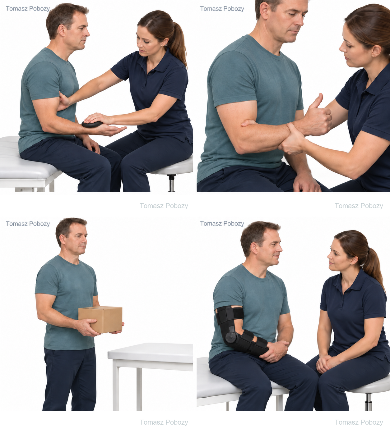
Illustrations show the rehabilitation stage. The treating team determines the movement range, brace settings and exercise dosage.
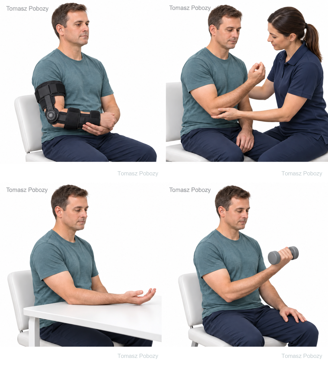
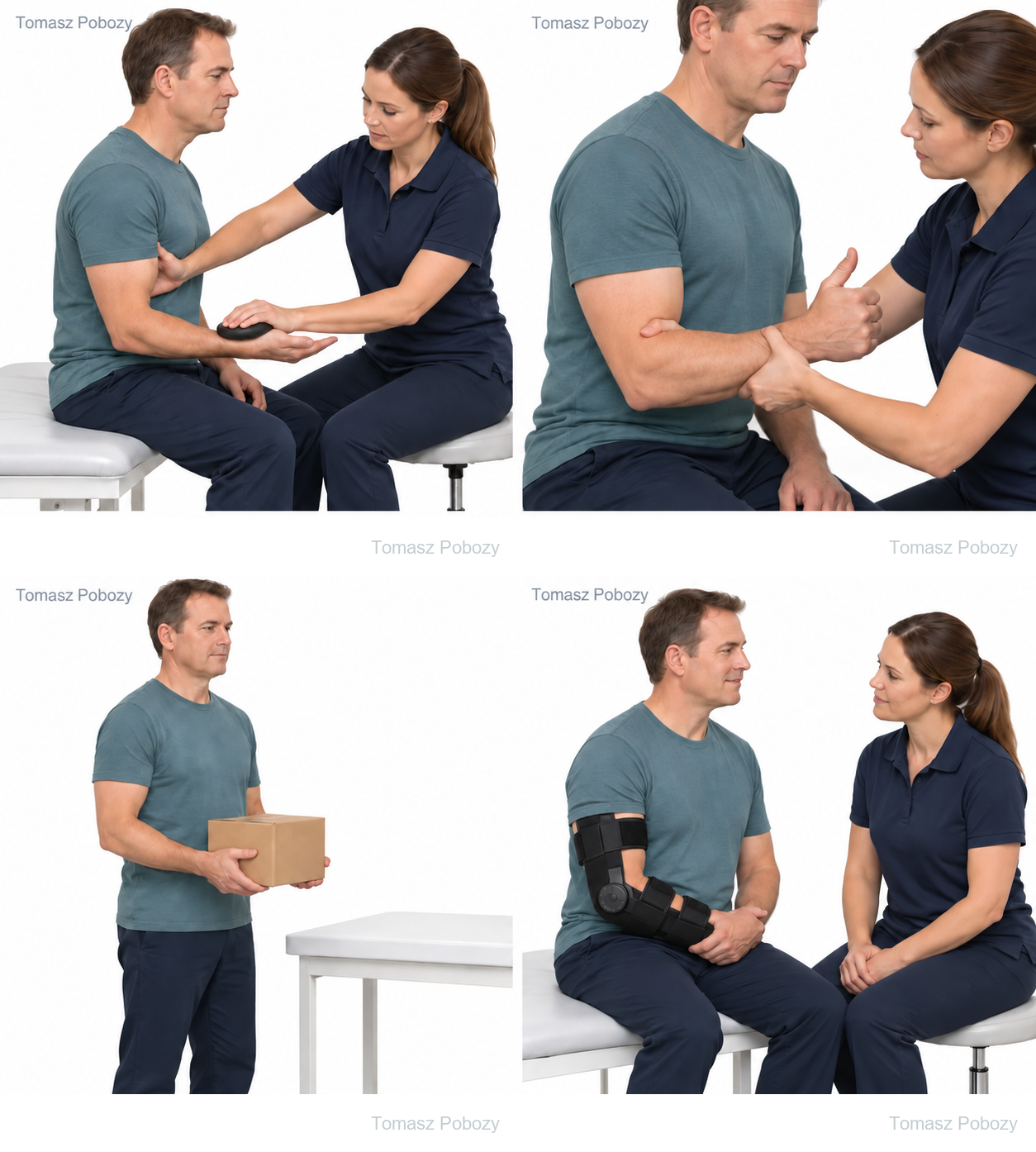
BOARD 02
Return criteria and safety
Strength and readiness
Assess pain-free motion, tolerance of progressive loads and work- or sport-specific tasks. Once testing is authorised, compare flexion and supination strength bilaterally. Logan’s commentary proposes a deficit below 10% as a supporting criterion. This is not a validated safety threshold; Phelps’s review found no established return-to-sport criteria. [1] [6]
Graded return to activity
A 2025 review reported a mean return to sport of 6.3 months. This describes study populations, not a mandatory deadline. Assess desk work, heavy manual work and sport separately; unrestricted return requires clinical clearance. [7]
Stop and arrange assessment
A sudden pop with pain, new bruising or loss of function; wound discharge, increasing redness or fever; or new weakness of finger or thumb extension require urgent contact with the surgical team. A cold, pale hand or rapidly increasing severe pain with swelling requires immediate assessment. Progressive loss of forearm rotation also needs investigation. [8]
For a life-threatening emergency, call your local emergency number immediately — 112 in the European Union. On the boards, 112 refers to the EU.
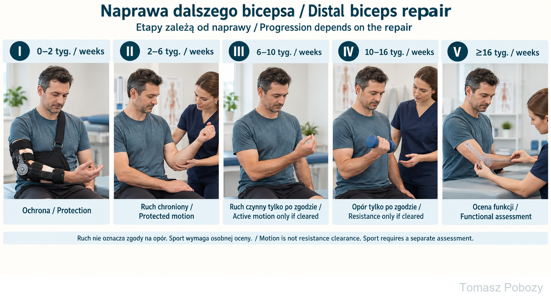
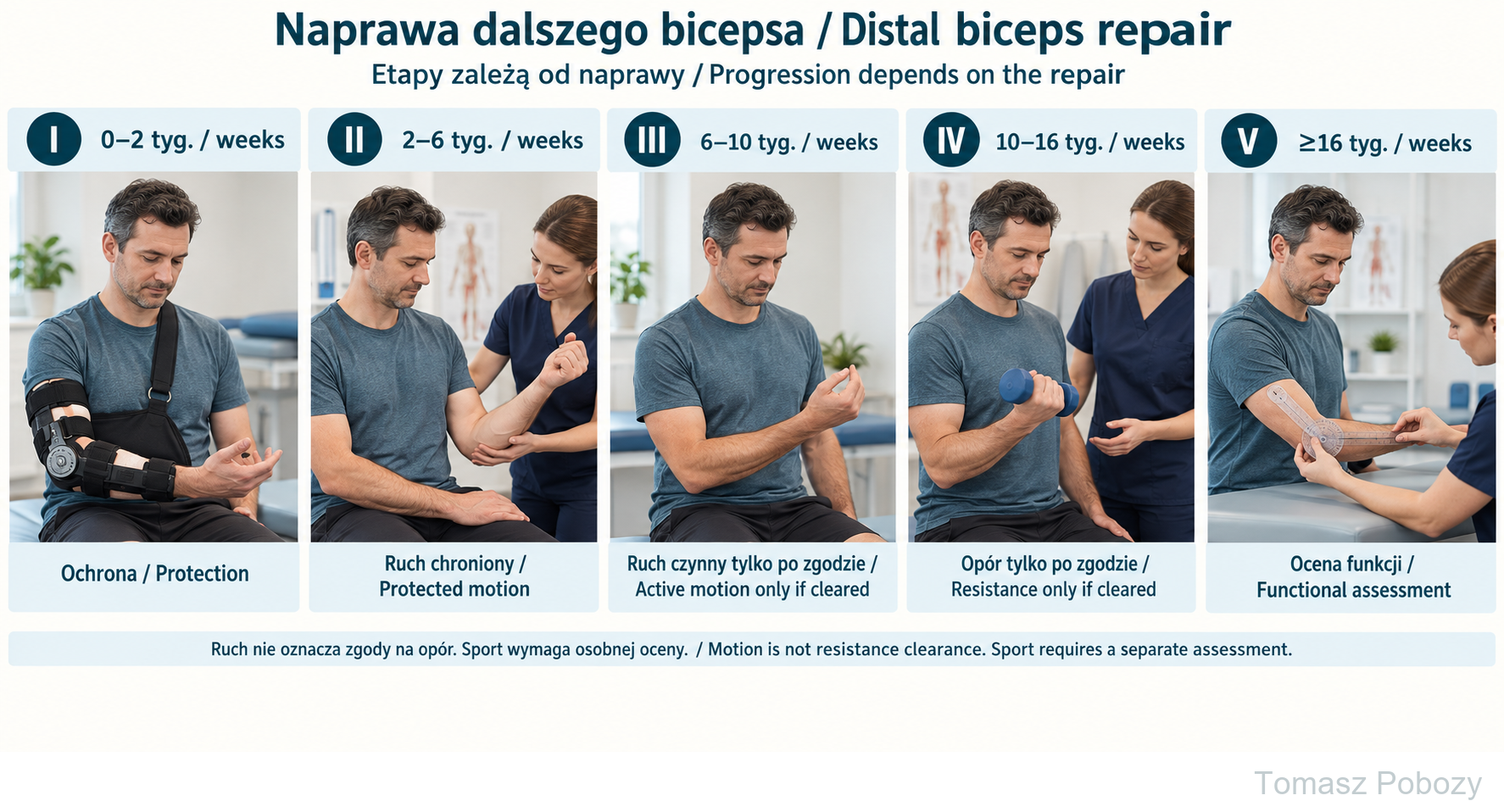
Brace protection; protected motion; active motion only after clearance; later low resistance; functional assessment. Timings match the page stages, not a universal prescription. More protective pathways may delay active motion or resistance. Measuring motion does not replace strength and sport-readiness assessment.
Exercises — a closer look
Use the complete boards above together with the instructions for the chosen stage. An exercise shown for a later stage is not permitted early.
Practising protected movement
Brace protection; protected motion; active motion only after clearance; later low resistance; functional assessment. Timings match the page stages, not a universal prescription. More protective pathways may delay active motion or resistance. Measuring motion does not replace strength and sport-readiness assessment.
Scope and protection
Before the first session
Progressing to active movement
After wound and swelling assessment, increase assisted and then active movement as instructed. Bergman’s RCT studied primary cortical-button repair: earlier active movement provided no clinically important advantage over 6 weeks of immobilisation. This does not justify early strength training. The surgeon determines when bracing can stop. [2]
Progressing to resistance
Published programmes differ substantially. Rogalski et al described strengthening from week 6 with a 2 kg (conservative rounded ceiling) limit until week 10 in a retrospective study. For all-suture anchor repair, Moroski et al propose isometrics from week 8, isotonics from week 10 and eccentrics from weeks 12–14; this is a technical report. Logan et al’s clinical commentary also delays active flexion and supination until approximately week 12; biceps isometrics start at week 12 and isotonics at week 16. Do not average these dates or transfer a weight limit between programmes. Agree a plan appropriate to the actual repair with the surgeon. Isometrics also load the attachment; pain, swelling or loss of range after exercise require reassessment. [1] [3] [4]
Additional scope notes
Rehabilitation protocol
The foundation is scientific literature. These stages provide a clinical synthesis, not a single programme validated in a comparative trial. The strongest randomised evidence concerns mobilisation timing after cortical-button fixation [2]. Detailed loading schedules come from observational research, a technical report and a clinical commentary [1] [3] [4]. Meta-analyses do not identify one clearly superior mobilisation strategy [5] [9]; certainty in the 2026 review was low or very low. Absence of a significant difference does not prove that all progressions are equivalent or that any early loading programme is safe.
Literature review: 21 September 2026. Full texts: [1,3–6]; abstracts and bibliographic records: [2,7–9].
For doctors and physiotherapists. Individualise this protocol according to the operative report, clinical examination and response to loading. The surgeon’s instructions take priority.
Prepared by Tomasz Poboży, MD, PhD — orthopaedic surgeon
References
References
- Logan CA, Shahien A, Haber D, Foster Z, Farrington A, Provencher MT. Rehabilitation Following Distal Biceps Repair. Int J Sports Phys Ther. 2019;14(2):308–317. DOI: 10.26603/ijspt20190308.
- Bergman JW et al. Is Immobilization Necessary for Early Return to Work Following Distal Biceps Repair Using a Cortical Button Technique? A Randomized Controlled Trial. J Bone Joint Surg Am. 2021;103(19):1763–1771. DOI: 10.2106/JBJS.20.02047.
- Rogalski BL et al. Outpatient versus At-Home Physical Therapy Following Distal Biceps Repair. Arch Bone Jt Surg. 2025;13(7):420–425. DOI: 10.22038/abjs.2025.81641.3717.
- Moroski N, Eskew JR, Marston G, Martin S. Distal Biceps Repair Using an All-Suture Anchor Technique. Arthrosc Tech. 2024;13(2):102841. DOI: 10.1016/j.eats.2023.09.016.
- Schönweger F et al. Distal biceps tendon repair: immobilization versus early mobilization. A systematic review and meta-analysis. EFORT Open Rev. 2026;11(7):849–858. DOI: 10.1530/EOR-2025-0152.
- Phelps BM et al. Postoperative rehabilitation and return to sport criteria following distal biceps tendon rupture surgery. JSES Int. 2025;9(3):929–933. DOI: 10.1016/j.jseint.2025.02.013.
- Boufadel P et al. Return to Sport After Distal Biceps Tendon Repair: A Systematic Review. Am J Sports Med. 2025;53(7):1769–1778. DOI: 10.1177/03635465241295618.
- Cain RA, Nydick JA, Stein MI, Williams BD, Polikandriotis JA, Hess AV. Complications following distal biceps repair. J Hand Surg Am. 2012;37(10):2112–2117. DOI: 10.1016/j.jhsa.2012.06.022.
- Simpson ER et al. No Clinically Significant Differences in Patient-Reported Outcomes and Range of Motion Between Early and Delayed Mobilization After Primary Distal Biceps Tendon Repair: A Systematic Review and Meta-analysis. Am J Sports Med. 2025;53(13):3261–3274. DOI: 10.1177/03635465251317207.
References and links checked on 18 October 2026. Existence of a publication does not validate every dose or timetable. Institutional protocols and examples require individualisation.
Use of this protocol
© 2026 Tomasz Poboży – orthopaedic surgeon. All rights reserved.