 Skip to content
FOR CLINICIANS AND PHYSIOTHERAPISTS
Elbow arthroscopy — rehabilitation protocol
Protection, movement and a gradual return to daily activity. Select the stage appropriate to the actual procedure and current clearance.
Using and loading the limb
Early movement does not permit lifting. Begin with light self-care without support, hanging or forceful twisting; resistance follows assessment.
Full guidance ↓Brace or immobilisation
A comfort sling is usually used for 24–72 hours and removed for movement. Splinting follows a prescription; this applies to isolated surgery without additional repair.
Full guidance ↓Permitted movement
Use early active and assisted motion without forceful overpressure. No routine 90° flexion limit; consider the extent of resection or release.
Full guidance ↓Permitted movement, loading and removal of a brace are separate decisions. Additional repairs may require a more protective programme.
Rehabilitation at a glance
Progression follows healing and functional criteria, not dates alone.
Rehabilitation boards
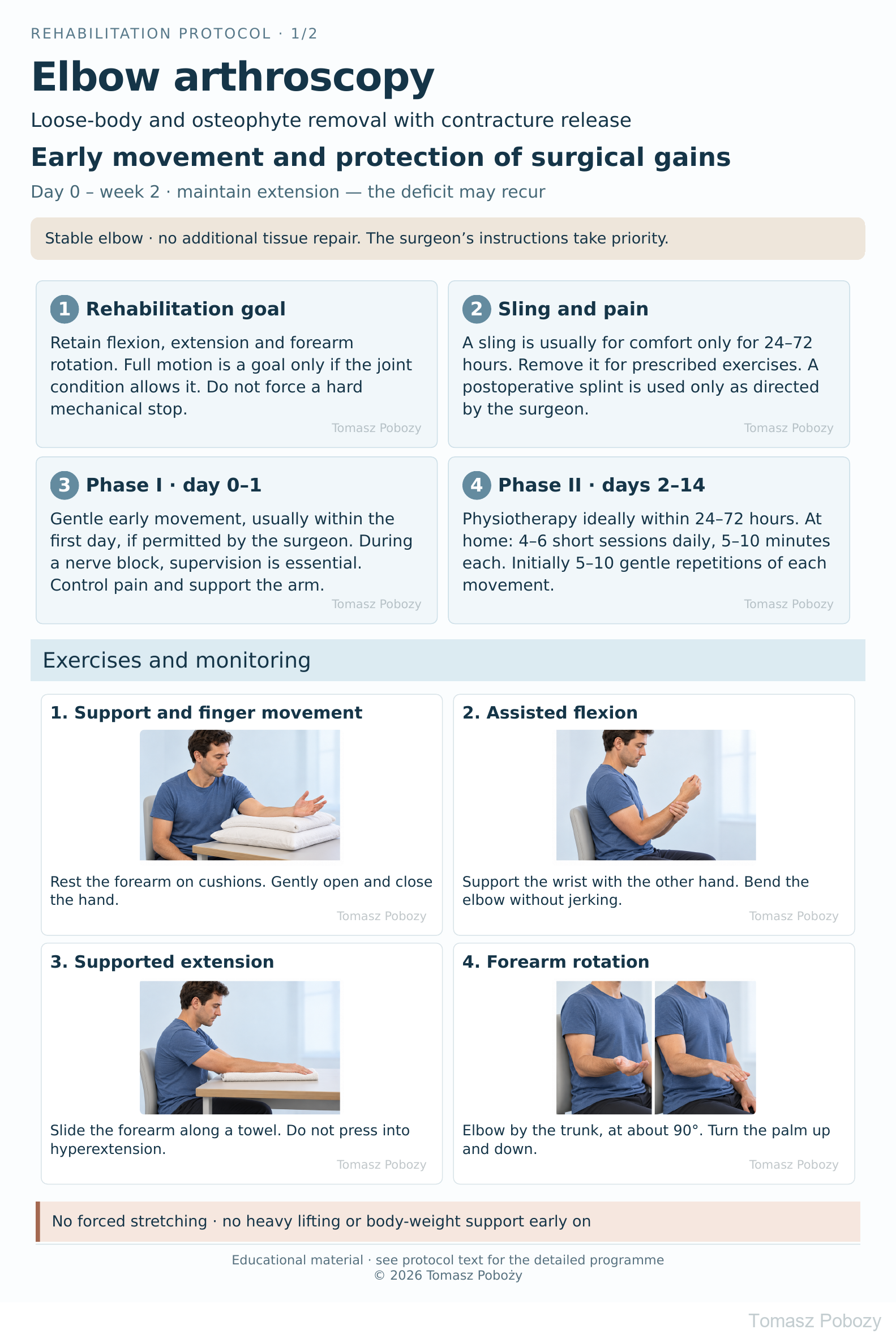
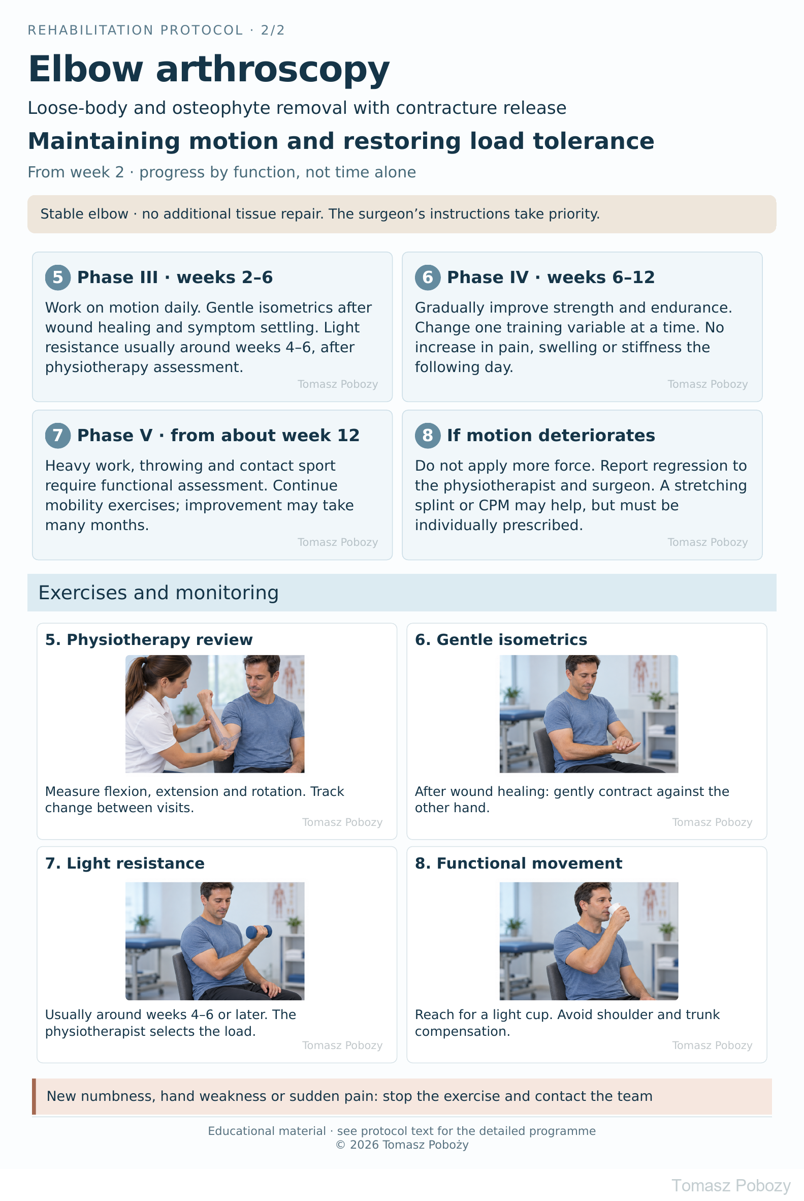
Exercises — a closer look
Use the complete boards above together with the instructions for the chosen stage. An exercise shown for a later stage is not permitted early.
Open the relevant complete board and zoom in. The viewer retains all stages and captions.
How should the illustrated exercises be performed?
Early exercises — board 1
- Support and fingers: rest the whole forearm on cushions without pressure in the elbow crease. Open and close the hand and move the wrist. Avoid forceful ball squeezing.
- Assisted flexion: keep the upper arm by the trunk and the shoulder relaxed. Support the wrist with the other hand and help bend the elbow. Avoid jerking or pressure on the wound.
- Supported extension: slide the forearm along a towel on a table and gently straighten the elbow. Adjust table height to avoid shoulder hiking. Do not press into hyperextension.
- Pronation and supination: keep the elbow by the trunk at about 90°. Rotate the forearm to turn the palm up and down. Do not substitute wrist deviation or whole-trunk rotation.
Later exercises — board 2
- Motion review: the physiotherapist measures flexion, extension deficit and rotation in a consistent position. Record pain, swelling and function, not just the best range immediately after mobilisation.
- Isometrics: with the elbow comfortably bent, gently contract against the other hand without moving. For example, five contractions of five seconds without holding the breath. The physiotherapist selects the direction of resistance.
- Light resistance: use an individually selected small weight or band, moving slowly without shoulder swinging. Do not add weight if motion decreases.
- Functional tasks: reaching for an empty cup, eating and dressing. Gradually increase range and task duration without abruptly increasing load.
Dosage and joint response
Initial early dose: 5–10 repetitions in 4–6 short sessions daily. After wound healing, the physiotherapist may introduce gentle stretching: 3–5 holds of 10–20 seconds without sharp pain. These are proposed starting doses for individual modification.
A mild stretching sensation is acceptable, but sharp pain, numbness or locking is not. If symptoms clearly increase after a session or the joint is stiffer and more swollen the next morning, reduce the dose and have the programme reviewed. Do not exercise harder simply to reach a particular angle.
Scope and protection
Scope, movement precautions and loading
Who is this for?
Following arthroscopic loose-body removal, osteophyte resection and contracture release, when the elbow is stable. Removing a mechanical block does not restore damaged cartilage or guarantee full motion.
This protocol does not cover ligament repair or reconstruction, biceps or triceps repair, fracture fixation, microfracture or other cartilage-restoration procedures. Following ulnar nerve decompression or transposition, permitted motion, wound protection and flexion tolerance must be confirmed.
Movement and sling use
Early mobilisation and avoiding prolonged immobilisation underpin rehabilitation [1],[2]. Proposed approach: a sling for comfort only, usually for 24–72 hours, with regular removal for prescribed exercises. If the orthopaedic surgeon prescribes a splint, its position and removal schedule take priority.
There is no routine 90° flexion limit. Work towards the range achievable after surgery without aggressive end-range pressure. Do not independently force or stretch a numb limb while a nerve block remains active.
Loading is a separate decision
Early movement does not mean lifting is permitted. Initially, light self-care is allowed within tolerance. Avoid heavy shopping bags, pushing up from a chair through the operated arm, push-ups, hanging and forceful twisting. After extensive bone resection, loading limits are set by the surgeon.
Stages and progression criteria
Phases I–III: surgery to week 6
Phase I — day 0–1
Goal: control pain and swelling and begin movement safely. Gentle flexion, extension and forearm rotation usually begin within the first day unless the surgeon advises otherwise. Initial instruction should take place before discharge. During a nerve block, movement requires team supervision; do not stretch independently.
Progression: the patient understands the exercises, pain allows participation, the dressing is not constrictive, and circulation and sensation are satisfactory. Severe pain despite treatment or an expanding haematoma requires reassessment.
Phase II — days 2–14
Goal: retain motion and allow wound healing. Physiotherapy ideally starts within 24–72 hours. Initially, 2–3 visits per week are proposed, more frequently if stiffness returns rapidly or exercises are difficult. At home, perform 4–6 sessions daily, 5–10 minutes each; start with 5–10 gentle repetitions of each movement.
The physiotherapist teaches active and assisted movement, selects gentle mobilisation and monitors joint response. Avoid forceful manipulation or painful stretching. Wound and suture review is usually around days 10–14, as specified at discharge.
Progression: the wound is healing well, swelling is settling, exercise does not cause a sustained flare, and motion is not deteriorating between visits. Loss of motion, increasing warmth or new nerve symptoms require review.
Phase III — weeks 2–6
Goal: further improve motion and restore muscle control. Continue daily exercise; the physiotherapist may add gentle end-range holds and scar treatment once the wound is fully closed. Gentle isometrics can begin after wound healing and symptom settling, often around weeks 2–3. Light resistance usually starts around weeks 4–6, only with stable motion and good tolerance.
Progression: no reactive swelling, tolerance of light tasks, and no meaningful deterioration the following day. If loading causes pain or stiffness, reduce it and return to the last well-tolerated level.
If your elbow becomes harder to straighten from one day to the next, contact your physiotherapist or surgeon early. Do not wait for the scheduled review or try to regain extension by applying increasing force.
Phases IV–V: from week 6 and return to activity
Phase IV — weeks 6–12
Goal: strength, endurance and task performance. Progressively strengthen elbow flexors and extensors, forearm muscles, grip and shoulder girdle. Perform resistance training 2–3 times weekly with a rest day between sessions. Start with 1–2 sets of 8–12 easy repetitions. Increase one variable at a time without loss of motion the following day.
Begin weight-bearing exercises only with the treating clinician’s approval, starting with light loading against a wall, not floor push-ups. Heavy work and sport are not automatically permitted at six weeks.
Phase V — from about week 12
Goal: resume demanding activities and consolidate recovery. Introduce work and sports tasks in stages. Throwing, racket sports, contact sports and heavy loads require assessment by the surgeon and physiotherapist. Return may be later after extensive contracture release.
Criteria: healed tissues, a stable range sufficient for the task, no pain or swelling after a loading trial, adequate strength and control, and no nerve symptoms. Full extension is not an absolute requirement if a persistent deficit is accepted by the surgeon. The week number does not replace an examination.
Mobility work may continue for many months [2]. Recurrent locking, progressive motion loss or increasing pain requires reassessment, not stronger stretching.
Daily activities and follow-up
Daily activities follow the permitted joint motion, lifting, grip and arm support for your procedure. Ending immobilisation is not clearance for lifting or driving. The detailed guidance below explains the criteria and exceptions.
Sling, rest, work, driving and adjunctive treatment
Rest: support the forearm and hand, avoid leaving the arm hanging for prolonged periods or resting the inner elbow on a hard surface. Do not keep the elbow bent all day. Night positioning or any splint is selected by the treating clinician.
Hygiene: manage the dressing as instructed at discharge. Do not soak the wound or use a swimming pool until it has fully healed. Do not massage fresh arthroscopy portals.
Work: light desk work is possible when pain allows short periods and the arm is supported. Take movement breaks. Manual work requires graded tasks and assessment of loading demands.
Driving: resume only without a sling, without medication impairing performance, and once safe steering and emergency manoeuvres are possible. There is no single safe date for everyone.
Stretching splints
Consider a splint for persistent or recurrent motion loss. A static-progressive stretching splint is different from protective immobilisation. Direction, force and wearing time are selected by the surgeon and physiotherapist; it is not routinely prescribed to everyone [2],[5]. Finger numbness, increased pain or hand colour changes require discontinuation and assessment.
CPM — continuous passive motion
CPM may be an adjunct after contracture release. Randomised trials found benefits in motion recovery, but do not establish a single universal regimen [3],[4]. Settings and duration require prescription; CPM does not replace learning independent exercises or clinical review.
Warning signs
Pain management and warning signs
Use medication as prescribed at discharge. Time exercises for effective pain relief, but do not use the absence of pain to force movement. Cooling through a cloth for about 10–15 minutes may help symptoms; do not apply to numb skin or directly over the wound. Check the skin.
Heterotopic ossification prophylaxis is considered by the surgeon according to risk and contraindications. This protocol does not recommend independently starting NSAIDs or a specific prophylaxis regimen [5].
Urgent same-day assessment
- Fever with increasing redness, warmth or wound discharge.
- New or worsening numbness, particularly in the little and ring fingers, or reduced grip and finger movement.
- Severe pain despite treatment, rapidly increasing swelling, haematoma or painful locking.
Immediate emergency care
A cold, pale or blue hand; rapidly increasing pain with tense swelling; sudden major movement weakness; breathlessness or chest pain: seek emergency care (112 / emergency department). Do not wait for a scheduled appointment.
Persistent burning, touch sensitivity, temperature or colour changes and disproportionate pain require early review for complex regional pain syndrome.
For a life-threatening emergency, call your local emergency number immediately — 112 in the European Union. On the boards, 112 refers to the EU.
Additional scope notes
Rehabilitation following arthroscopic joint debridement and contracture release. The priorities are to retain the motion gained during surgery, control pain and swelling, and gradually restore function.
From the first days after surgery, the priority is to maintain the range of motion gained during the operation. Practise elbow bending and straightening regularly, paying particular attention to extension: an extension deficit may recur after contracture release. Perform short, frequent sessions as instructed by your physiotherapist, without forceful pressure or pushing through pain. Do not neglect flexion or forearm rotation.
References
References
- Verstuyft L, Caekebeke P, van Riet R. Postoperative rehabilitation in elbow surgery. J Clin Orthop Trauma. 2021;20:101479. DOI: 10.1016/j.jcot.2021.101479.
- Siemensma MF, van der Windt AE, van Es EM, Colaris JW, Eygendaal D. Management of the stiff elbow: a literature review. EFORT Open Rev. 2023;8(5):351–360. DOI: 10.1530/EOR-23-0039.
- O’Driscoll SW, Rojas Lievano J, Morrey ME, et al. Prospective Randomized Trial of Continuous Passive Motion Versus Physical Therapy After Arthroscopic Release of Elbow Contracture. J Bone Joint Surg Am. 2022;104(5):430–440. DOI: 10.2106/JBJS.21.00685.
- Huang L, Wang Y, Ma Y, Ding J, Lu S. Does Early Continuous Passive Motion Improves Effectiveness of Physical Therapy After Arthroscopic Release of Elbow Contracture: A Prospective Randomized Trial. Am J Phys Med Rehabil. 2025;104(5):445–451. DOI: 10.1097/PHM.0000000000002646.
- Schneider MM, Rentschler V, Geyer S, et al. Rehabilitation after surgical release of the stiff elbow: A literature review. J Orthop. 2025;64:51–58. DOI: 10.1016/j.jor.2024.10.061.
References and links checked on 18 October 2026. Existence of a publication does not validate every dose or timetable. Institutional protocols and examples require individualisation.
Clinical commentary and terms of use
Commentary
Early movement, not aggressive stretching
After loose-body and osteophyte removal with contracture release, rehabilitation should use the newly available motion without sustaining inflammation through excessive loading. Restrictions from ligament reconstruction or arthroplasty protocols should not be transferred to this procedure.
Surgeon and physiotherapist
The physiotherapist should receive the operative report: extent of resection, achieved motion, stability assessment, cartilage status, ulnar nerve management and restrictions. Record preoperative motion, the first rehabilitation assessment, then regular measurements during the early weeks and at reviews around weeks 6 and 12. Regression warrants earlier contact with the surgeon.
Strength of evidence
No single postoperative regimen is validated for everyone [5]. The exact session frequency, repetitions and strengthening dates here form a proposed clinical programme, not the direct findings of one trial. CPM trials support considering this treatment, not making it mandatory for every patient [3],[4].