 Skip to content
ORTHOPAEDIC DIAGNOSTICS
Ankle Ultrasound — When Is It Useful and What Can It Miss?
Ankle ultrasound is particularly useful for questions involving tendons, selected ligaments, tendon sheaths, effusion or other superficial soft tissues. It does not automatically replace X-rays, MRI or CT. Selection depends on injury mechanism, symptom duration and the suspected structure.
When is ultrasound particularly useful?
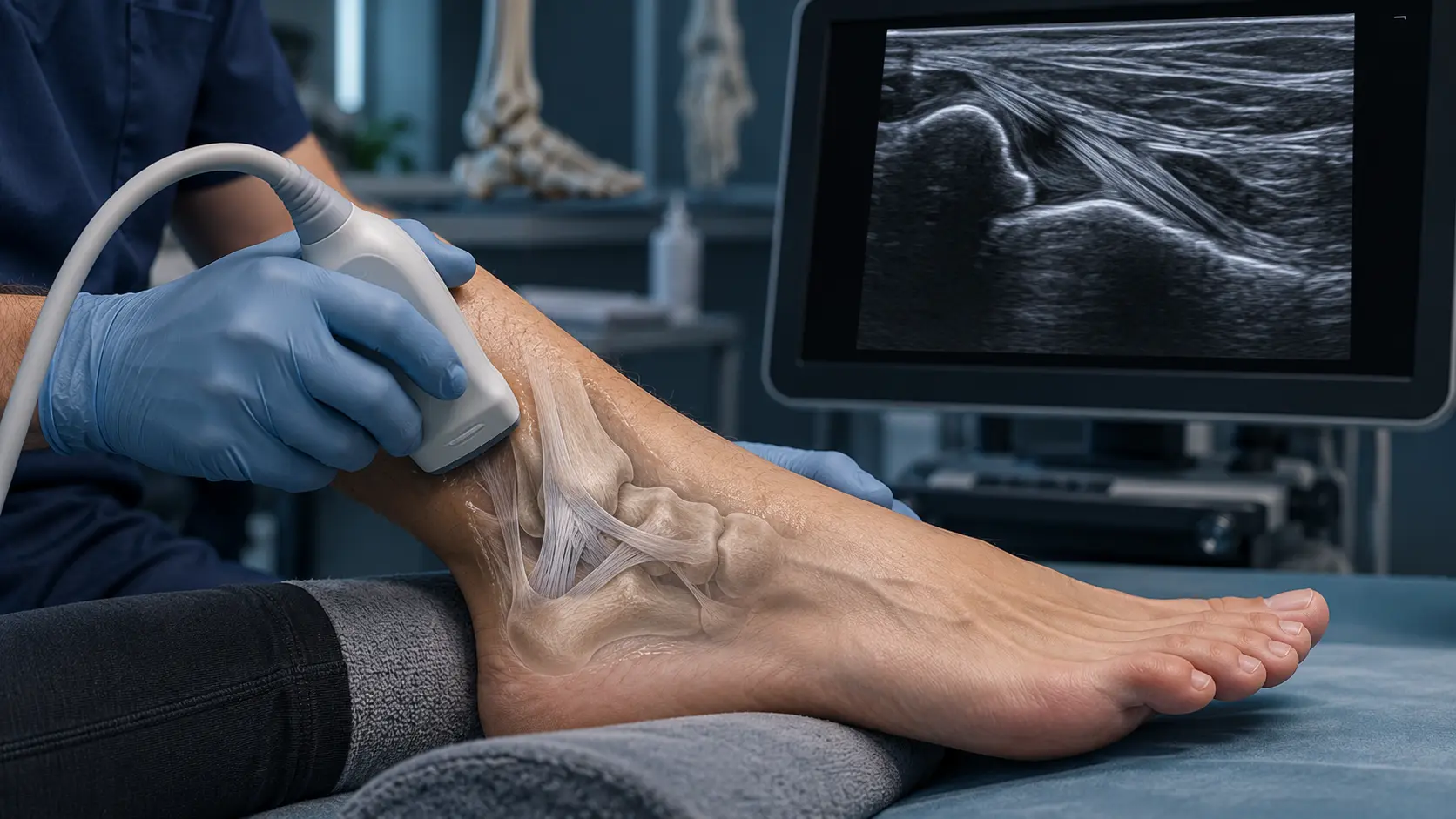
Ultrasound assesses soft tissues in real time and, for selected problems, during movement. Common uses include suspected lateral ankle ligament injury, Achilles tear or tendinopathy, peroneal tendon problems, posterior tibial tendon pathology, tenosynovitis and selected superficial periarticular lesions.
After an ankle sprain: ultrasound or X-rays first?
After acute injury, the first task is to assess fracture risk. When examination and the Ottawa rules indicate this possibility, X-rays are the appropriate initial test. Ultrasound is not routinely the first investigation for every acute ankle injury. It can be valuable as a targeted test when a particular superficial structure, especially a tendon or selected ligament, needs closer assessment.
More about test selection after a sprain: Sprained Ankle — When Are X-rays, Ultrasound or MRI Needed? (in Polish)
Chronic pain: ultrasound is not always first
For chronic ankle pain of uncertain cause, investigation often begins with X-rays. If examination suggests a particular tendon, ultrasound or MRI may be appropriate next. Cartilage, osteochondral lesions, intra-articular impingement, bone or deeper structures more often require MRI and, selectively, CT.
Can ultrasound show ligament injury?
It can be highly accurate for selected lateral ligaments, particularly the anterior talofibular ligament. Meta-analyses confirm high diagnostic accuracy for this ligament. This does not imply equal reliability for all ankle ligaments: assessment depends on the structure, injury phase and examiner experience. Findings must be interpreted with the mechanism and examination.
Can ultrasound show an Achilles tendon tear?
Ultrasound is well suited to Achilles assessment, showing structure, thickness, partial or complete discontinuity and tendinopathic changes. A sudden snap, loss of push-off strength or suspected acute rupture requires prompt clinical examination; ultrasound can confirm and characterise the injury.
When is MRI or CT better?
MRI may be preferable for the whole joint and deeper structures, especially suspected osteochondral lesions, cartilage injury, marrow pathology or complex trauma. CT is particularly useful when detailed bone assessment is central.
What happens during the examination?
Several positions are used to access the anterior, lateral, medial and posterior ankle. Depending on the problem, the clinician may ask you to move the foot or perform gentle dynamic tests. Previous X-rays, MRI or CT should be compared with clinical and ultrasound findings where available.
Questions to discuss with an orthopaedic surgeon
Does ultrasound replace MRI?
No. They have different strengths. Ultrasound is useful for many tendons, selected ligaments and superficial structures, and allows dynamic examination. MRI better shows bone marrow, cartilage and deeper joint structures.
Should ultrasound be performed immediately after a sprain?
Not always. First assess the injury clinically and decide whether X-rays are needed. Ultrasound is most useful when there is a specific soft-tissue question and the result may change management.
References
- American College of Radiology. ACR Appropriateness Criteria®: Chronic Ankle Pain. Current imaging selection criteria.
- American College of Radiology. ACR Appropriateness Criteria®: Acute Trauma to the Ankle. Imaging selection after acute injury and the Ottawa rules.
- Kocsis K, Stubnya B, Váncsa S, et al. Diagnostic accuracy of ultrasonography in acute lateral ankle ligament injury: A systematic review and meta-analysis. Injury. 2024;55(Suppl 3):111730. doi:10.1016/j.injury.2024.111730.
Related resources
- Orthopaedic diagnosis
- Sprained Ankle — When Are X-rays, Ultrasound or MRI Needed? (in Polish)
- Ankle injuries
- Foot and Ankle Conditions
Medical author: Tomasz Poboży - orthopaedic surgeon, MD, PhD, specialist in orthopaedics and musculoskeletal traumatology. Educational material does not replace examination or individually selected investigations. Updated: 12 September 2026.