 Skip to content
FOR CLINICIANS AND PHYSIOTHERAPISTS
Intramuscular and submuscular ulnar nerve transposition — postoperative protocol
Protection, movement and a gradual return to daily activity. Select the stage appropriate to the actual procedure and current clearance.
Using and loading the limb
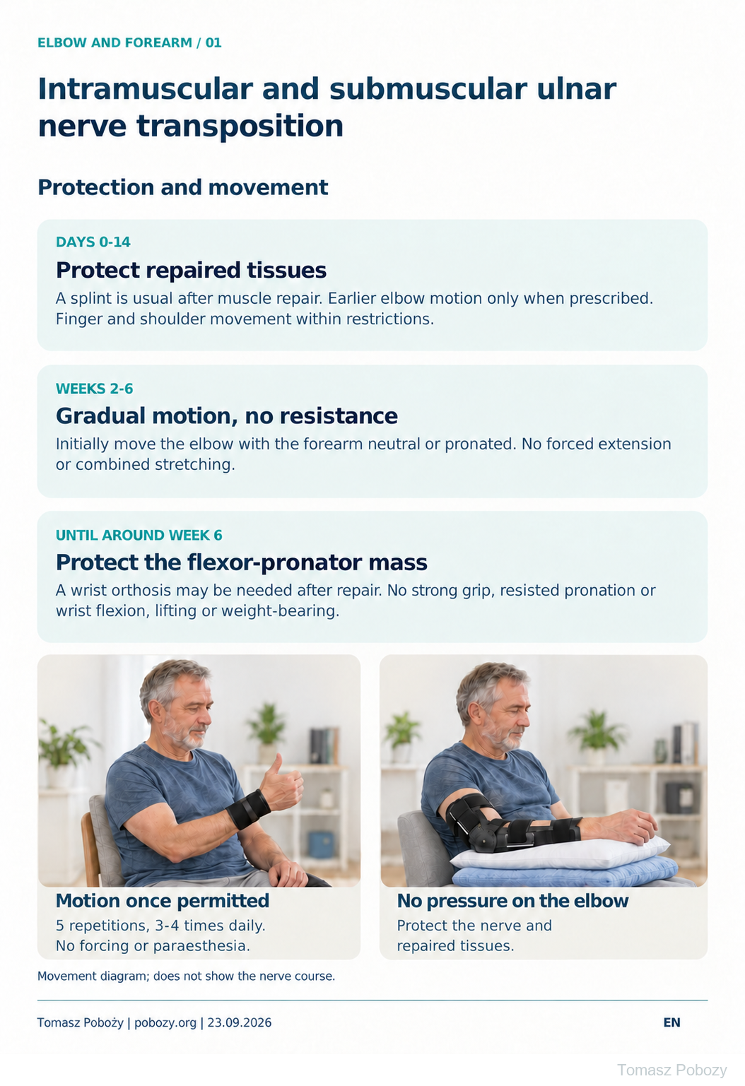
After flexor-pronator repair, avoid resisted pronation, wrist flexion, forceful grip, lifting and body-weight support through week 6.
Full guidance ↓Brace or immobilisation
After muscle-mass repair, a surgeon-set splint is usually used for 10–14 days; later wrist bracing may be needed. Do not remove protection for movement without clearance.
Full guidance ↓Permitted movement
Introduce movement gradually. Initially avoid combining end-range elbow extension, supination and wrist extension.
Full guidance ↓Permitted movement, loading and removal of a brace are separate decisions. Additional repairs may require a more protective programme.
Rehabilitation at a glance
Progression follows healing and functional criteria, not dates alone.
Rehabilitation boards
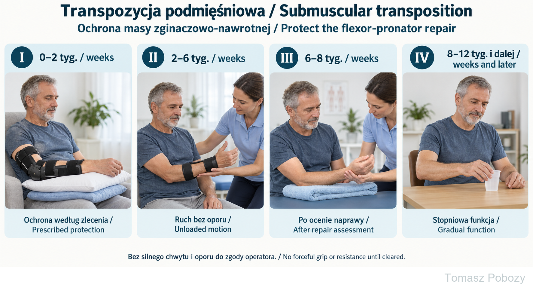
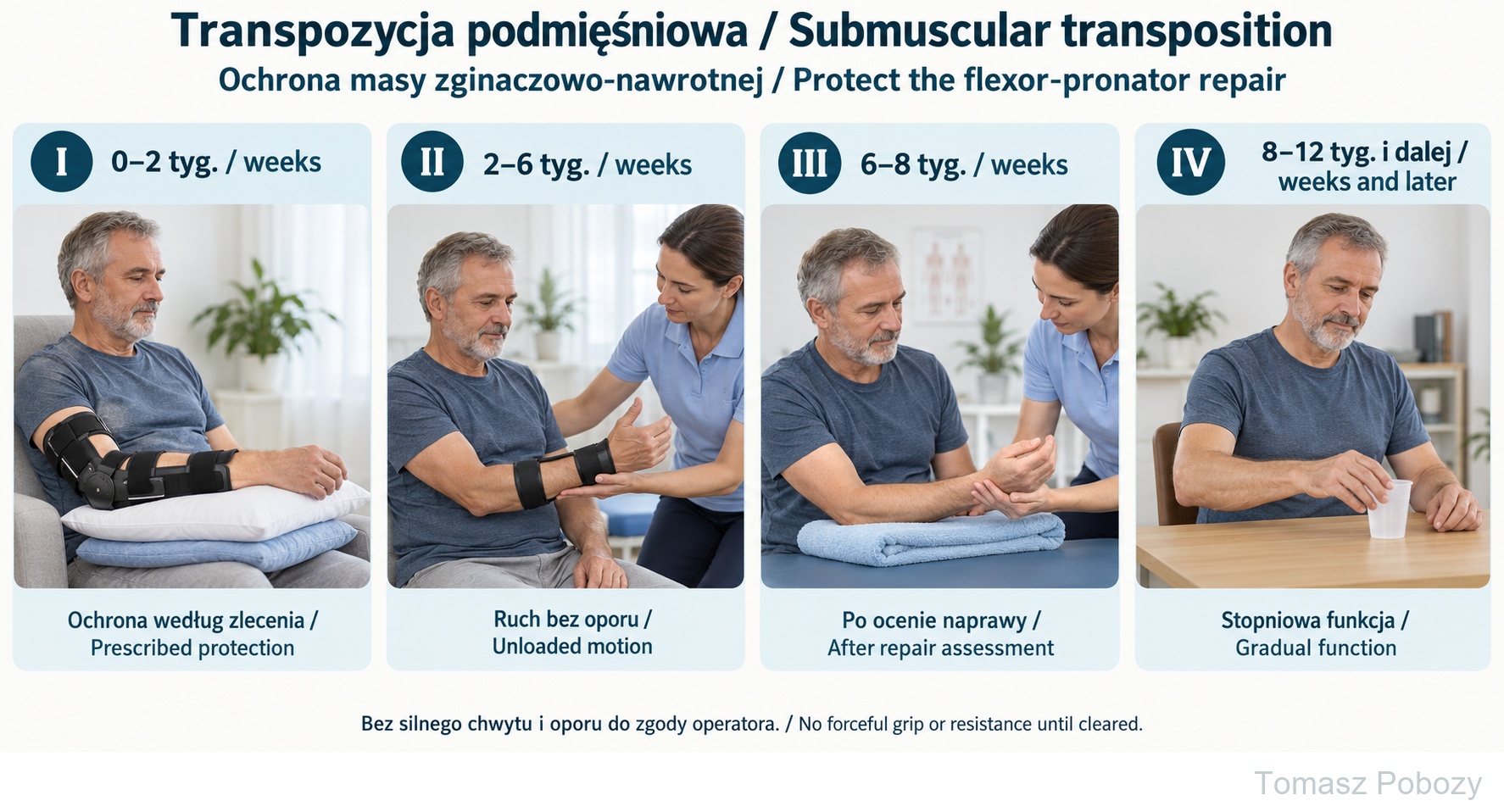
Weeks 0–2: protection appropriate for the repair. Weeks 2–6: permitted unloaded motion. Weeks 6–8: healing assessment before light strengthening. Weeks 8–12 and later: light tasks progressed by tolerance. Splint angle and design are individual.
Exercises — a closer look
Use the complete boards above together with the instructions for the chosen stage. An exercise shown for a later stage is not permitted early.
Open the relevant complete board and zoom in. The viewer retains all stages and captions.
Exercise instructions and progression are contained in the stage descriptions linked above.
Weeks 0–2: protection appropriate for the repair. Weeks 2–6: permitted unloaded motion. Weeks 6–8: healing assessment before light strengthening. Weeks 8–12 and later: light tasks progressed by tolerance. Splint angle and design are individual.
Scope and protection
Scope and protection pathway
This protocol covers primary anterior intramuscular or submuscular ulnar nerve transposition. The shared pathway protects a divided, lengthened or reattached flexor-pronator mass. It excludes nerve repair or grafting, revision surgery, ligament reconstruction and fracture fixation.
Review the operative report: extent of muscle dissection, repair method and nerve stability. If the origin was not repaired, the orthopaedic surgeon may shorten protection and permit earlier movement. The label “intramuscular” alone does not justify accelerated rehabilitation.
Exercises and daily protection
Start with 5 comfortable elbow movements, 3–4 times daily, within permitted limits. Gently open and close the hand and spread and bring together the fingers. No forceful squeezing, exercise balls, weights or resistance bands. Adjust dosage to swelling, pain and sensory symptoms.
Until the end of week 6, avoid resisted pronation, wrist flexion and forceful grip, including isometrics that load the repair. No lifting loads, pushing, pulling or weight-bearing through the operated limb. Light self-care is permitted within protective restrictions.
Avoid pressure on the medial elbow and aggressive nerve mobilisation. Gentle nerve sliders require individual instruction and must not increase paraesthesia. Begin scar care only after complete wound closure; avoid deep pressure over the nerve.
Sport and recreation
Walking from the first days as tolerated. Lower-limb training must avoid gripping, weight-bearing or loading the operated arm and avoid fall risk.
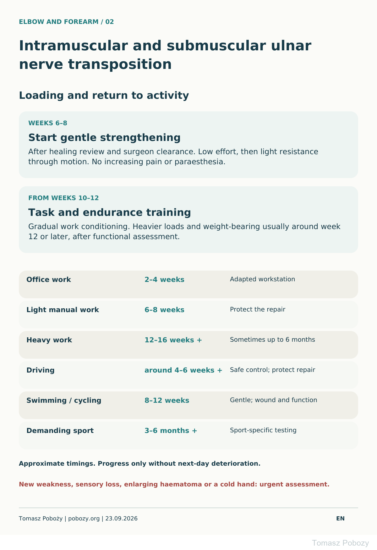
Gentle swimming and outdoor cycling are usually considered from weeks 8–12: a healed wound, functional motion and good exercise tolerance are required. Cycling requires reliable steering and braking. Vigorous swimming is treated as sport training.
Heavy gym training, climbing, throwing, racquet and contact sports: gradual return usually during months 3–6 or later. Completion of strength progression and sport-specific testing is required. Time alone does not provide clearance.
Progression criteria and therapy
Healed wound, no increasing swelling, adequate task-specific motion, stable sensation and hand control. Before loading, assess repair-site tenderness, strength and endurance, then review the response through the following day.
Hand therapy supports orthosis fitting, motion recovery and monitoring of sensation, intrinsic strength and dexterity. Lack of motion progress by weeks 4–6 needs review; do not independently increase stretching force. Additional treatments for established stiffness require repair assessment first.
Follow-up and nerve recovery
Reviews are usually at 10–14 days and around weeks 6 and 12, more often for wound problems, stiffness or neurological deficits. Document little-finger and ulnar ring-finger sensation, finger movement, elbow motion and loading tolerance.
Muscle and fascial healing and restored motion do not mean nerve recovery is complete. Sensory and strength recovery takes months; final results may be assessed at 12–18 months. Advanced neuropathy with muscle wasting may recover incompletely. New deterioration requires investigation regardless of time since surgery.
Stages and progression criteria
Days 0–14: dressing and immobilisation
After flexor-pronator repair, use a long-arm splint as prescribed, usually for 10–14 days. The elbow is commonly flexed approximately 60–90°, with the wrist neutral or slightly flexed; the surgeon determines exact forearm and joint positioning.
Keep the dressing clean and dry. Support the limb and elevate the hand for swelling. A sling provides comfort and protection during mobility. Protect the anaesthetised arm from injury and temperature extremes. Gently move the fingers and the shoulder within restrictions. Do not remove the splint for elbow exercises without permission.
Earlier controlled motion requires explicit surgeon instructions and does not authorise loading. Wound review and removal of non-absorbable sutures are usually at 10–14 days.
Weeks 2–6: gradual movement
After review, start gentle active or active-assisted elbow motion, initially with the forearm neutral or pronated. Increase the arc gradually without forcing extension. Introduce supination with the elbow flexed according to tolerance and surgeon instructions.
Initially avoid combining end-range elbow extension, supination and wrist extension. Exercise joints separately without sustained stretching. After origin repair, a wrist orthosis may be prescribed until around week 6; its position and removal for exercises are individualised.
Gentle active wrist motion is usually introduced during weeks 3–4 after clearance, avoiding end-range or sustained extension. Discontinue or convert the elbow splint to a removable orthosis according to repair stability; six weeks of complete elbow immobilisation is not automatic.
Weeks 6–8: transition to strengthening
After healing assessment and surgeon clearance, gradually discontinue wrist protection, increase motion and introduce light resistance. Begin with submaximal isometrics, followed by low resistance through movement. Progress particularly cautiously when loading the repaired flexor-pronator mass.
Initial dosage: one short set of 5–10 repetitions on alternate days without maximal effort. Persistent origin tenderness or nerve irritation requires postponing load increases. Do not apply a universal weight limit independently of repair quality.
Weeks 8–12 and beyond
Develop grip, wrist, forearm, elbow and shoulder strength and endurance. Increase one demand at a time: repetition tolerance first, then resistance and task difficulty.
Consider work conditioning from around weeks 10–12. Weight-bearing and heavier loads are generally introduced around week 12 or later, after surgeon clearance and functional testing. Pain, swelling or paraesthesia persisting the next day requires reduced dosage.
Daily activities and follow-up
Daily activities follow the permitted joint motion, lifting, grip and arm support for your procedure. Ending immobilisation is not clearance for lifting or driving. The detailed guidance below explains the criteria and exceptions.
Work and driving
Office work approximately 2–4 weeks with adapted duties and workstation. Support the forearm without elbow pressure and take frequent breaks. Light manual work usually 6–8 weeks; heavy work often 12–16 weeks, sometimes up to 6 months depending on repair and residual deficits.
Assess driving individually, usually around weeks 4–6 or later. Requirements include no restrictive splint or sling, secure grip and full vehicle control, and an emergency manoeuvre without pain or compromising repair protection. Do not drive while taking medication that impairs performance.
Warning signs
Symptoms requiring assessment
Urgent contact: new grip or finger weakness, worsening sensation after the anaesthetic wears off, an enlarging haematoma, substantial swelling or increasing pain. A cold, pale or blue hand requires immediate care.
Fever, spreading redness, drainage or wound separation require review. Sudden medial elbow pain after loading with loss of function may indicate repair disruption. Painful snapping, disproportionate pain and hand temperature or colour changes also require assessment.
For a life-threatening emergency, call your local emergency number immediately — 112 in the European Union. On the boards, 112 refers to the EU.
Additional scope notes
Protect the nerve and musculofascial repair. Motion according to repair stability. Strengthening usually from weeks 6–8.
For physicians and physiotherapists. Restrictions and progression require patient assessment, the operative report and surgeon instructions. Timings are approximate and do not replace functional assessment.
References
References
- BayCare Clinic. Ulnar Nerve Submuscular Transposition. Rehabilitation protocol. Updated 2021. Source
- Bienz T. Rehabilitation Guidelines: Cubital Tunnel Release – Submuscular. The Hand Rehabilitation Center of Indiana. Source
- Hand and Upper Limb Centre. Ulnar Nerve Transposition: Post-Operative Protocol. January 2019. Source
- Washington University in St. Louis. Transmuscular Ulnar Nerve Transposition. Surgical Education. Source
- So SP, Lee WS, Ku K, Shin YH, Kim JK. Modified musculofascial lengthening technique for submuscular ulnar nerve transposition in cubital tunnel syndrome. PLoS One. 2025;20(1):e0318303. doi:10.1371/journal.pone.0318303. Source
- American Academy of Orthopaedic Surgeons. Cubital Tunnel Release. OrthoInfo. Source
- Gateshead Health NHS Foundation Trust. Cubital Tunnel Release Surgery. 2024. Source
References and links checked on 18 October 2026. Existence of a publication does not validate every dose or timetable. Institutional protocols and examples require individualisation.
Use of this protocol
© 2026 Tomasz Poboży – orthopaedic surgeon · pobozy.org