 Skip to content
FOR CLINICIANS AND PHYSIOTHERAPISTS
Distal humerus fracture — rehabilitation protocol
Protection, movement and a gradual return to daily activity. Select the stage appropriate to the actual procedure and current clearance.
Using and loading the limb
No lifting, pushing, pulling or support until healing review. Week 6 alone does not permit resistance; greater loads require confirmed union.
Full guidance ↓Brace or immobilisation
Splint or sling according to stability and wound, usually short term. A hinged brace is not mandatory after every stable fixation.
Full guidance ↓Permitted movement
Distinguish intact triceps, repaired triceps and olecranon osteotomy. Active extension after osteotomy/repair requires explicit clearance.
Full guidance ↓Permitted movement, loading and removal of a brace are separate decisions. Additional repairs may require a more protective programme.
Rehabilitation at a glance
Progression follows healing and functional criteria, not dates alone.
Rehabilitation boards
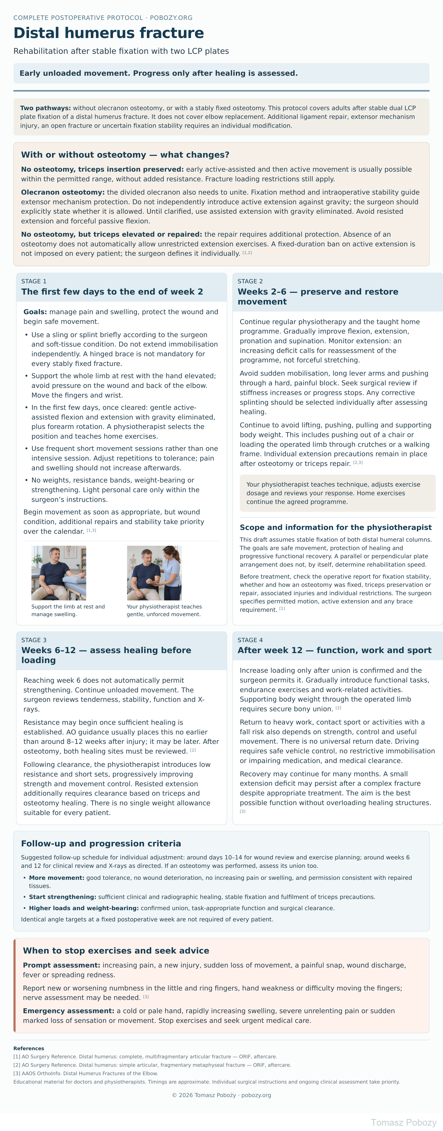
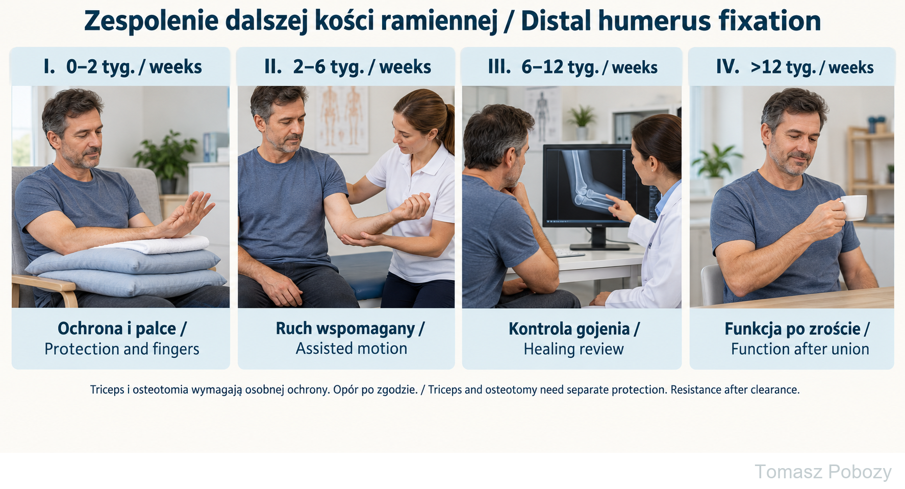
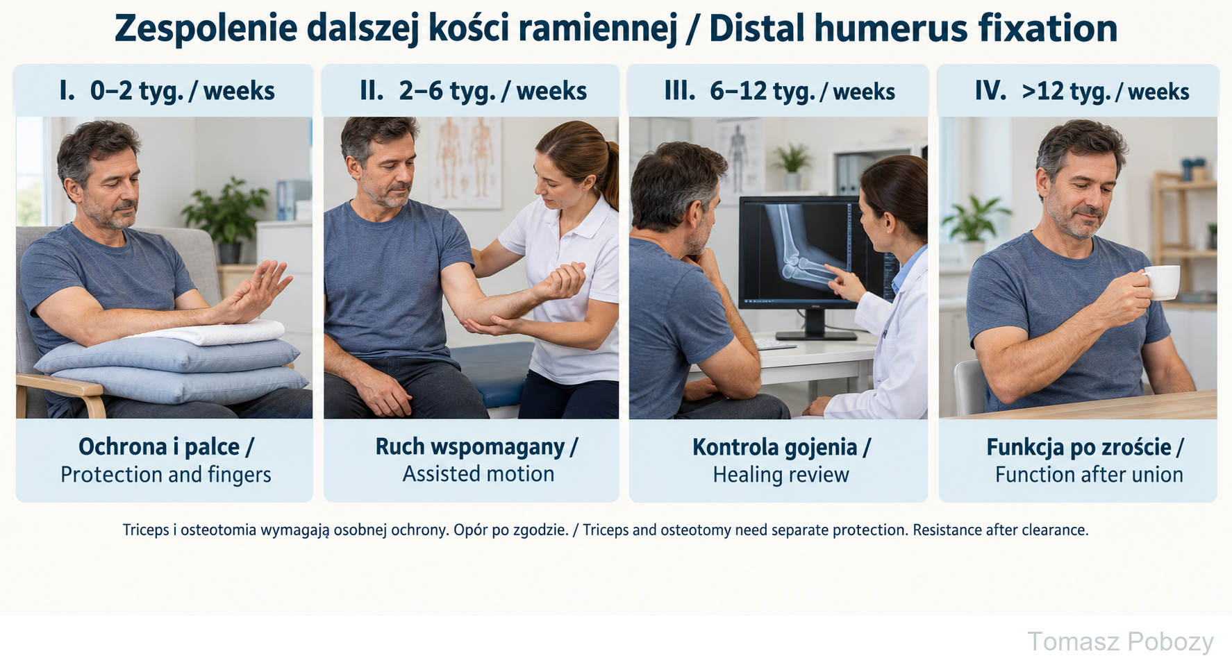
Weeks 0–2: support and finger motion. Weeks 2–6: permitted assisted motion. Weeks 6–12: healing review before resistance, usually no earlier than weeks 8–12 after assessment. After week 12: gradual function only with sufficient union. Osteotomy or triceps repair requires separate extension precautions. The consultation is illustrative, not a patient radiograph.
Exercises — a closer look
Use the complete boards above together with the instructions for the chosen stage. An exercise shown for a later stage is not permitted early.
Open the relevant complete board and zoom in. The viewer retains all stages and captions.
Exercise instructions and progression are contained in the stage descriptions linked above.
Weeks 0–2: support and finger motion. Weeks 2–6: permitted assisted motion. Weeks 6–12: healing review before resistance, usually no earlier than weeks 8–12 after assessment. After week 12: gradual function only with sufficient union. Osteotomy or triceps repair requires separate extension precautions. The consultation is illustrative, not a patient radiograph.
Scope and protection
1. Scope and information for the physiotherapist
This protocol assumes stable fixation of both distal humeral columns. The goals are safe movement, protection of healing and progressive functional recovery. A parallel or perpendicular plate arrangement does not, by itself, determine rehabilitation speed.
Before treatment, check the operative report for fixation stability, whether and how an osteotomy was fixed, triceps preservation or repair, associated injuries and individual restrictions. The surgeon specifies permitted motion, active extension and any brace requirement. [1]
2. With or without osteotomy — what changes?
No osteotomy, triceps insertion preserved: early active-assisted and then active movement is usually possible within the permitted range, without added resistance. Fracture loading restrictions still apply.
Olecranon osteotomy: the divided olecranon also needs to unite. Fixation method and intraoperative stability guide extensor mechanism protection. Do not independently introduce active extension against gravity; the surgeon should explicitly state whether it is allowed. Until clarified, use assisted extension with gravity eliminated. Avoid resisted extension and forceful passive flexion.
No osteotomy, but triceps elevated or repaired: the repair requires additional protection. Absence of an osteotomy does not automatically allow unrestricted extension exercises. A fixed-duration ban on active extension is not imposed on every patient; the surgeon defines it individually. [1],[2]
7. Follow-up and progression criteria
Suggested follow-up schedule for individual adjustment: around days 10–14 for wound review and exercise planning; around weeks 6 and 12 for clinical review and X-rays as directed. If an osteotomy was performed, assess its union too.
- More movement: good tolerance, no wound deterioration, no increasing pain or swelling, and permission consistent with repaired tissues.
- Start strengthening: sufficient clinical and radiographic healing, stable fixation and fulfilment of triceps precautions.
- Higher loads and weight-bearing: confirmed union, task-appropriate function and surgical clearance.
Identical angle targets at a fixed postoperative week are not required of every patient.
Stages and progression criteria
3. The first few days to the end of week 2
Goals: manage pain and swelling, protect the wound and begin safe movement.
- Use a sling or splint briefly according to the surgeon and soft-tissue condition. Do not extend immobilisation independently. A hinged brace is not mandatory for every stably fixed fracture.
- Support the whole limb at rest with the hand elevated; avoid pressure on the wound and back of the elbow. Move the fingers and wrist.
- In the first few days, once cleared: gentle active-assisted flexion and extension with gravity eliminated, plus forearm rotation. A physiotherapist selects the position and teaches home exercises.
- Use frequent short movement sessions rather than one intensive session. Adjust repetitions to tolerance; pain and swelling should not increase afterwards.
- No weights, resistance bands, weight-bearing or strengthening. Light personal care only within the surgeon’s instructions.
Begin movement as soon as appropriate, but wound condition, additional repairs and stability take priority over the calendar. [1],[3]
4. Weeks 2–6 — preserve and restore movement
Continue regular physiotherapy and the taught home programme. Gradually improve flexion, extension, pronation and supination. Monitor extension: an increasing deficit calls for reassessment of the programme, not forceful stretching.
Avoid sudden mobilisation, long lever arms and pushing through a hard, painful block. Seek surgical review if stiffness increases or progress stops. Any corrective splinting should be selected individually after assessing healing.
Continue to avoid lifting, pushing, pulling and supporting body weight. This includes pushing out of a chair or loading the operated limb through crutches or a walking frame. Individual extension precautions remain in place after osteotomy or triceps repair. [2],[3]
5. Weeks 6–12 — assess healing before loading
Reaching week 6 does not automatically permit strengthening. Continue unloaded movement. The surgeon reviews tenderness, stability, function and X-rays.
Resistance may begin once sufficient healing is established. AO guidance usually places this no earlier than around 8–12 weeks after injury; it may be later. After osteotomy, both healing sites must be reviewed. [2]
Following clearance, the physiotherapist introduces low resistance and short sets, progressively improving strength and movement control. Resisted extension additionally requires clearance based on triceps and osteotomy healing. There is no single weight allowance suitable for every patient.
6. After week 12 — function, work and sport
Increase loading only after union is confirmed and the surgeon permits it. Gradually introduce functional tasks, endurance exercises and work-related activities. Supporting body weight through the operated limb requires secure bony union. [2]
Return to heavy work, contact sport or activities with a fall risk also depends on strength, control and useful movement. There is no universal return date. Driving requires safe vehicle control, no restrictive immobilisation or impairing medication, and medical clearance.
Recovery may continue for many months. A small extension deficit may persist after a complex fracture despite appropriate treatment. The aim is the best possible function without overloading healing structures. [3]
Warning signs
8. When to stop exercises and seek advice
Prompt assessment: increasing pain, a new injury, sudden loss of movement, a painful snap, wound discharge, fever or spreading redness.
Report new or worsening numbness in the little and ring fingers, hand weakness or difficulty moving the fingers; nerve assessment may be needed. [3]
Emergency assessment: a cold or pale hand, rapidly increasing swelling, severe unrelenting pain or sudden marked loss of sensation or movement. Stop exercises and seek urgent medical care.
For a life-threatening emergency, call your local emergency number immediately — 112 in the European Union. On the boards, 112 refers to the EU.
Additional scope notes
Early, regular movement helps limit elbow stiffness. Stable fixation permits unloaded exercises but does not replace bone healing. Work with a physiotherapist within the movement and tissue protection limits specified by your orthopaedic surgeon.
References
References
- AO Surgery Reference. ORIF — Plate fixation. Complete and multifragmentary articular fracture of the distal humerus. Aftercare.
- AO Surgery Reference. ORIF — Plate fixation. Complete articular fracture, simple articular, fragmentary metaphyseal. Aftercare.
- AAOS. Distal Humerus Fractures of the Elbow. OrthoInfo.
References and links checked on 18 October 2026. Existence of a publication does not validate every dose or timetable. Institutional protocols and examples require individualisation.