 Skip to content
FOR CLINICIANS AND PHYSIOTHERAPISTS
Dupuytren contracture surgery — rehabilitation after limited fasciectomy
Protection, movement and a gradual return to daily activity. Select the stage appropriate to the actual procedure and current clearance.
Using and loading the limb
Protect skin and wound early; avoid forceful grip and forced extension. Strengthening follows skin closure and tolerance of light activities.
Full guidance ↓Brace or immobilisation
Night splinting is not routine for everyone. Consider it for progressive extension loss or other indications, with fit checks.
Full guidance ↓Permitted movement
Begin prescribed hand exercise early; guidance favours days 3–5 when the wound allows. Avoid independent forceful passive extension.
Full guidance ↓Permitted movement, loading and removal of a brace are separate decisions. Additional repairs may require a more protective programme.
Rehabilitation at a glance
Progression follows healing and functional criteria, not dates alone.
Rehabilitation boards

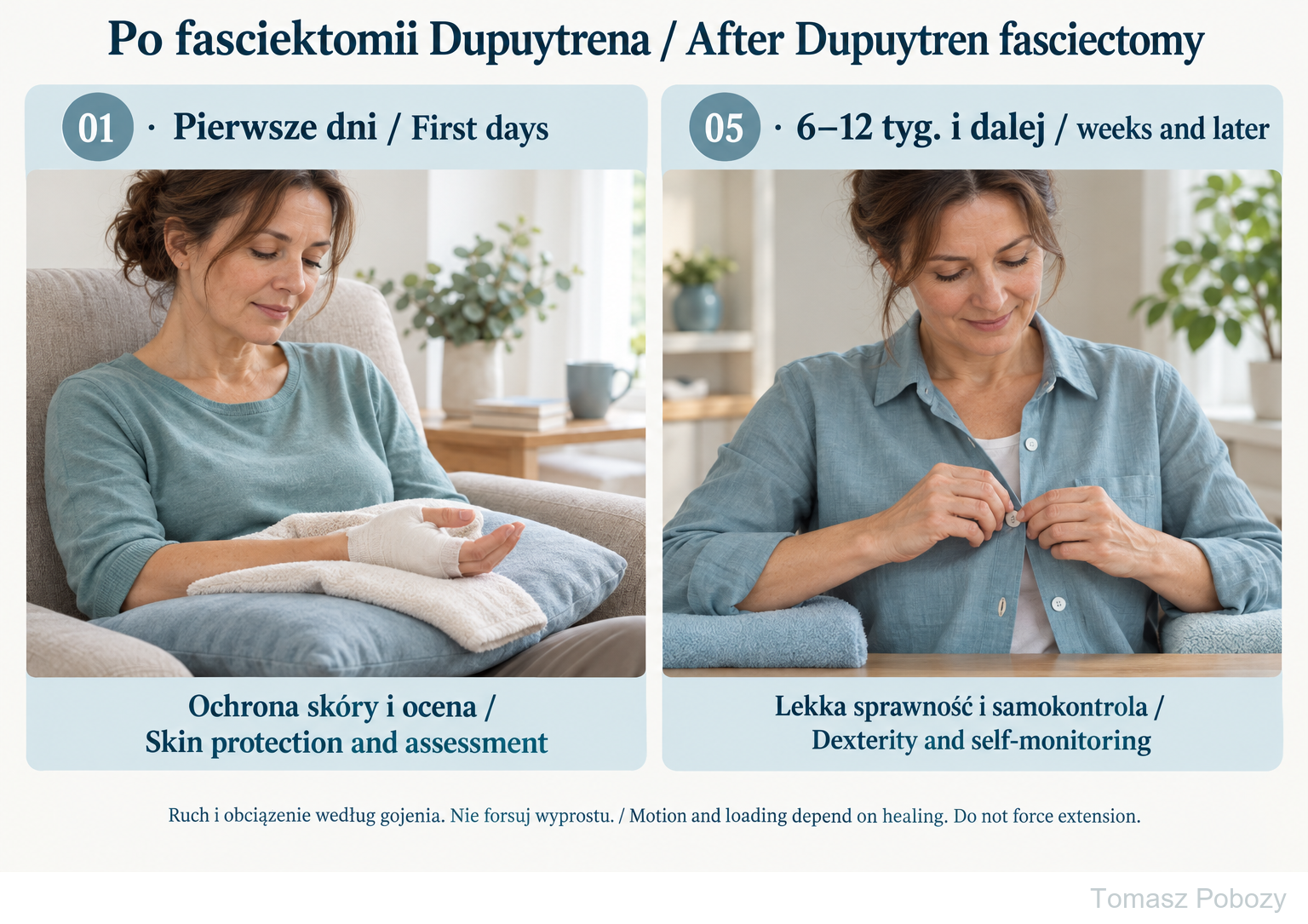
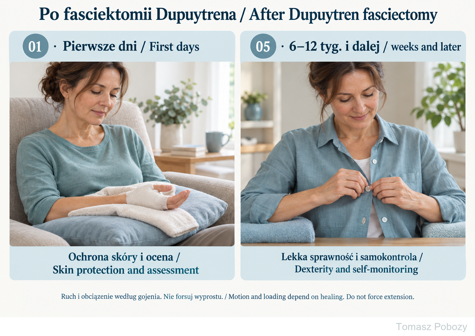
Phase 01: support, skin protection and assessment before starting the programme. Phase 05: light dexterity, such as fastening buttons, and monitoring the response. The full board illustrates phases 02–04: finger motion, hook posture and later light grip. Do not force extension or fist closure.
Exercises — a closer look
Use the complete boards above together with the instructions for the chosen stage. An exercise shown for a later stage is not permitted early.
Open the relevant complete board and zoom in. The viewer retains all stages and captions.
Exercise instructions and progression are contained in the stage descriptions linked above.
Phase 01: support, skin protection and assessment before starting the programme. Phase 05: light dexterity, such as fastening buttons, and monitoring the response. The full board illustrates phases 02–04: finger motion, hook posture and later light grip. Do not force extension or fist closure.
Scope and protection
Initial assessment and monitoring
Record the operated fingers, extent of excision and any additional procedures. Measure active and passive extension and flexion at the metacarpophalangeal (MCP) and proximal interphalangeal (PIP) joints; use the same method for serial measurements. Assess swelling, skin, sensation and tasks important to the patient. Test grip strength only when resistance is permitted. An unexplained difference between active and passive restriction calls for assessment of its cause, not an automatic increase in stretching force.
Orthosis: use according to indications
Do not routinely prescribe a night orthosis for every patient after fasciectomy [1,2]. Consider one for a progressive extension deficit, capsular flexion contracture or after arthrolysis. Position and low corrective force should be selected by the treating team. When an orthosis is indicated, the guideline describes night use for 6–24 weeks with regular review and adjustment [1]; this is not a compulsory duration for every patient. New pain, sensory disturbance, skin irritation or changes in finger colour require a fit check. Do not independently increase the extension force or modify a prescribed orthosis without agreement.
Stages and progression criteria
01–02 · First days and early movement
01 · First days: protection and assessment
From surgery until a safe movement programme is established
Goal: Limit swelling, protect the skin and identify restrictions arising from surgery.
Actions: Support the forearm comfortably and elevate the hand intermittently. Review the operative record, circulation, sensation, pain and swelling. Agree the permitted movement and any orthosis with the orthopaedic surgeon; move unrestricted joints of the limb.
Exercises: Do not independently start passive straightening of the operated fingers. Arrange early hand therapy; the guideline favours starting exercises on days 3–5 if local tissue condition and instructions permit [1].
Protection: No forceful gripping, weight-bearing through the hand or forced extension. A skin graft or additional reconstruction rules out automatic use of this schedule.
Progression and review: Progress when restrictions are clear, local tissue condition is stable and movement has been permitted. New sensory loss, increasing pain or a change in finger colour requires assessment, not forceful exercise.
02 · Early finger movement
Usually from days 3–5 to about week 2; once movement is permitted
Goal: Maintain the extension achieved and gradually recover flexion without overloading the skin.
Actions: Use short, gentle movement sessions. The guideline suggests 3–6 sessions daily [1]; tailor their duration and exercise selection to the hand’s response. Check pain, swelling and movement quality after each session.
Exercises: A · Support the forearm and keep the wrist comfortable. Slowly open the hand to the permitted extension, then bend the fingers into a loose partial fist. Do not push with the other hand. Example starting dose: 5 slow repetitions; reduce the range or number if skin tension or pain increases.
Protection: Do not force the fingertips to touch the palm or press the whole hand flat on a table. Avoid sudden stretching and resistance. Passive movement requires individual instruction first.
Progression and review: Progress when movement does not impair healing, symptoms return to baseline after sessions and the extension deficit is not increasing. Deterioration persisting into the next day calls for dose reduction and reassessment.
03 · Tendon gliding and dexterity
Approximately weeks 2–6; according to healing and tolerance
Goal: Improve independent finger joint movement and light grip during everyday activities.
Actions: Continue exercise A. Introduce short tasks such as moving light objects and fastening buttons. Gentle scar mobilisation may begin after complete skin closure and instruction; do not perform it over an unhealed surface.
Exercises: B · From straight fingers, bend the interphalangeal joints while keeping the knuckles at the finger bases as straight as comfortable: this forms a hook position. Return to permitted extension. Support the forearm and keep the wrist relaxed. Example: 5 cycles without resistance in selected movement sessions, if the therapist permits this position.
Protection: Select passive stretching and intrinsic muscle exercises after examination; do not add strong pressure. Avoid prolonged forceful gripping and rubbing a sensitive scar.
Progression and review: Before strengthening: completely closed skin, tolerance of light tasks, stable movement and no increasing pain or swelling. If extension is being lost, assess the cause and whether an orthosis is indicated.
04 · Gradual strengthening
Often around weeks 4–6 or later; only after meeting the criteria
Goal: Rebuild grip tolerance without aggravating the tissue response.
Actions: Once resistance is permitted, begin with a soft sponge and brief light tasks. Maintain movement exercises. This stage is not a mandatory date for starting forceful hand exercise.
Exercises: C · Support the forearm. Gently squeeze a soft sponge, then fully relax the grip. Do not squeeze maximally or hold your breath. Suggested starting dose: 5 repetitions, 2–3 sessions per week with a rest day between them.
Protection: Increase one variable at a time: repetitions, task duration or resistance. Do not immediately add heavy tools, forceful twisting or body-weight support.
Progression and review: Progress when the next session and following day bring no increased swelling, pain or loss of movement. If symptoms worsen, return to the previously tolerated dose and seek advice for persistent symptoms.
05 · Return to function and self-monitoring
Usually weeks 6–12 and beyond; according to activity demands
Goal: Recover the dexterity required and maintain the functional result.
Actions: Grade work, recreation and task practice. Example: start with 2–5 minutes of a light task, then pause to assess the hand. Set duration and resistance separately; heavy manual work requires assessment of grip, skin, sensation and safety.
Exercises: Continue the movement exercises still needed. The guideline describes a usual 3–12 weeks of therapy; stopping depends on scar stability and resolution of relevant functional and activity limitations [1], not the calendar alone.
Protection: Resume driving only with a reliable grip, steering control and the ability to respond safely in an emergency, without the effects of medicines that impair performance. There is no single date for every patient.
Progression and review: Persistent or increasing contracture, hypersensitivity and declining daily function require review. Not every loss of extension represents disease recurrence; do not decide this without examination.
Warning signs
Warning signs and reasons to change the programme
Urgent medical assessment: a pale, blue or cold finger; new or increasing loss of sensation; sudden pain or rapidly increasing swelling. Stop exercising and do not wait for the planned appointment. Fever, increasing redness, discharge or skin breakdown require prompt contact with the surgeon. Disproportionate pain, marked hypersensitivity, temperature or colour changes and progressive stiffness may indicate complex regional pain syndrome and need early investigation. Do not try to overcome stiffness by force.
For a life-threatening emergency, call your local emergency number immediately — 112 in the European Union. On the boards, 112 refers to the EU.
Additional scope notes
Protecting healing tissues, recovering finger movement and gradually returning to hand use. The complete illustrated guide presents the stages and exercise examples; expandable programme details follow below.
This programme applies to adults after partial (limited) fasciectomy with primary skin closure and without additional tendon, nerve or vascular repair. It is not a ready-to-use programme after dermofasciectomy with a skin graft, needle fasciotomy or collagenase treatment. Arthrolysis, revision surgery, impaired healing or circulation require modification by the treating team.
A specific case, a specific patient, a specific surgeon. Timings are reference points. Progression depends on skin condition, flexion and extension, swelling, sensation and load tolerance. Instructions arising from the operative findings take priority.
The example doses of 5 repetitions, short task sessions and 2–3 strengthening sessions per week are practical suggestions for individual adjustment, not doses validated in a comparative trial. Do not perform the entire set when only selected exercises have been prescribed.
References
References
- Kemler MA, de Wijn RS, van Rijssen AL et al. Dutch Multidisciplinary Guideline on Dupuytren Disease. J Hand Surg Glob Online. 2023;5(2):178–183. doi:10.1016/j.jhsg.2022.11.008
- Jerosch-Herold C, Shepstone L, Chojnowski AJ et al. Night-time splinting after fasciectomy or dermo-fasciectomy for Dupuytren’s contracture: a pragmatic, multi-centre, randomised controlled trial. BMC Musculoskelet Disord. 2011;12:136. doi:10.1186/1471-2474-12-136
- Lorenz FM, Henning E, Sicher C, Langner I. Effects of postoperative hand therapy in patients with Dupuytren’s disease: a prospective hyperspectral imaging study. Die Orthopädie. 2025;54:386–394. doi:10.1007/s00132-025-04631-w
References and links checked on 18 October 2026. Existence of a publication does not validate every dose or timetable. Institutional protocols and examples require individualisation.
Clinical commentary and terms of use
Commentary: surgeon–therapist cooperation
The goal is a useful, functional hand, not extension at any cost. The surgeon communicates details of the procedure and the tissues requiring protection. The physiotherapist or hand therapist assesses function, selects exercises, checks technique and adjusts the dose within the instructions. Feedback should cover loss of extension as well as difficulty bending, pain and everyday function. Incomplete extension after a severe contracture does not automatically justify aggressive stretching.
The strongest support is for avoiding routine night orthoses in all operated patients [2]. Detailed exercise recommendations in the guideline largely reflect consensus [1]. A 2025 study included 35 patients and short-term microcirculatory measures; it does not establish the optimal exercise dose or long-term functional outcome [3]. The stage windows and example doses are a practical programme proposal requiring patient assessment.