 Skip to content
FOR CLINICIANS AND PHYSIOTHERAPISTS
First MTP joint arthroplasty — rehabilitation protocol
Protection, movement and a gradual return to daily activity. Select the stage appropriate to the actual procedure and current clearance.
Using and loading the limb
Load as prescribed, usually in a stiff postoperative shoe. Initially avoid rolling through the great toe, heel raises and jumping.
Full guidance ↓Brace or immobilisation
Use a stiff shoe or rocker sole according to implant and wound, later wider footwear. This is an MTP I arthroplasty programme, not fusion rehabilitation.
Full guidance ↓Permitted movement
Begin gentle great-toe motion after clearance and wound assessment without forcing. Implant type and additional procedures matter.
Full guidance ↓Permitted movement, loading and removal of a brace are separate decisions. Additional repairs may require a more protective programme.
Rehabilitation at a glance
Progression follows healing and functional criteria, not dates alone.
Rehabilitation boards
Exercises — a closer look
Use the complete boards above together with the instructions for the chosen stage. An exercise shown for a later stage is not permitted early.
Open the relevant complete board and zoom in. The viewer retains all stages and captions.
How should the exercises shown on the poster be performed?
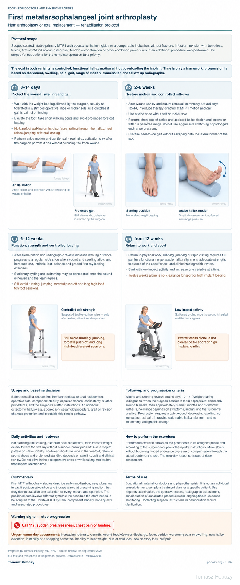
Perform the exercise shown on the poster only in its assigned phase and according to the surgeon’s or physiotherapist’s instructions. Move slowly, without bouncing, forced end-range pressure or compensation through the lateral border of the foot. The next-day response is part of dose assessment.
Scope and protection
Scope and baseline decision
Before rehabilitation, confirm: hemiarthroplasty or total replacement, operative side, component stability, capsular closure, cheilectomy or other procedures, and the surgeon’s written instructions. An additional osteotomy, hallux-valgus correction, sesamoid procedure, graft or revision changes protection and is outside this simple pathway.
Follow-up and progression criteria
Wound and swelling review: around days 10–14. Weight-bearing radiographs, when the surgeon considers them appropriate: commonly around 6 weeks, then approximately 3 and 6 months and 12 months; further surveillance depends on symptoms, implant and the surgeon’s practice. Progression requires a quiet wound, decreasing swelling, no increasing rest pain, improving gait, stable hallux alignment and no concerning radiographic change.
Stages and progression criteria
Phase 1 · 0–14 days — protect the wound, swelling and gait
Walk with the weight bearing allowed by the surgeon, usually as tolerated in a stiff postoperative shoe or rocker sole; use crutches if gait is painful or limping. Elevate the foot, take short walking bouts and avoid prolonged forefoot loading. No barefoot walking on hard surfaces, rolling through the hallux, heel raises, jumping or lateral loading. Perform ankle motion and gentle, pain-free hallux activation only after the surgeon permits it and without stressing the fresh wound.
Phase 2 · 2–6 weeks — restore motion and controlled roll-over
After wound review and suture removal, commonly around days 12–14, introduce therapy directed at MTP I motion and gait. Use a wide shoe with a stiff or rocker sole. Perform short sets of active and assisted hallux flexion and extension within a pain-free range; do not use aggressive stretching or prolonged end-range pressure. Practise heel-to-toe gait without escaping onto the lateral border of the foot.
Phase 3 · 6–12 weeks — function, strength and controlled loading
After examination and radiographic review, increase walking distance, progress to a regular wide shoe when wound and swelling allow, and introduce calf, intrinsic-foot, balance and graded first-ray loading exercises. Stationary cycling and swimming may be considered once the wound is healed and the team agrees. Still avoid running, jumping, forceful push-off and long high-load forefoot sessions.
Phase 4 · from 12 weeks — return to work and sport
Return to physical work, running, jumping or rapid cutting requires full painless functional range, stable hallux alignment, adequate strength, tolerance of the specific task and clinical/radiographic review. Start with low-impact activity and increase one variable at a time. Twelve weeks alone is not clearance for sport or high implant loading.
Daily activities and follow-up
Progress in everyday activities follows the permitted loading and motion for your procedure. Walking without aids requires a controlled gait; returning to driving requires safe vehicle control and no impairing medication. The detailed recommendations below explain the criteria and exceptions.
Daily activities and footwear
For standing and walking, establish heel contact first, then transfer weight calmly toward the first ray without a sudden hallux push-off. Use a step-to pattern on stairs initially. Footwear should be wide in the forefoot; return to sports shoes and prolonged standing depends on swelling, gait and clinical review. Do not drive in the postoperative shoe or while taking medication that impairs reaction time.
Warning signs
Call 112: sudden breathlessness, chest pain or fainting. New calf pain/swelling, wound breakdown, cold or blue toes or sudden loss of function require urgent same-day assessment.
Warning signs
Urgent review is needed for increasing redness, warmth, wound breakdown or discharge, fever, sudden worsening pain or swelling, new hallux deviation, instability or a snapping sensation, inability to bear weight, blue or cold toes, new sensory loss, calf pain. Stop progression and contact the team.
For a life-threatening emergency, call your local emergency number immediately — 112 in the European Union. On the boards, 112 refers to the EU.
Additional scope notes
A protocol for adults after primary first metatarsophalangeal arthroplasty with the Doratek/PIEX MTP I system distributed by MED&CARE. It covers hemiarthroplasty and total joint replacement when no additional osteotomy or reconstruction requires a separate pathway.
Scope: isolated, stable primary MTP I arthroplasty for hallux rigidus or a comparable indication, without fracture, infection, revision with bone loss, fusion, first-ray/Akin/Lapidus osteotomy, tendon reconstruction or other combined procedures. If an additional procedure was performed, the orthopaedic surgeon’s instructions for the complete operation take priority.
The goal in both variants is controlled, functional hallux motion without overloading the implant. Time is only a framework; progression is based on the wound, swelling, pain, gait, range of motion, examination and follow-up radiographs.
References
References
- Giza E, Sullivan M, Ocel D, et al. First metatarsophalangeal hemiarthroplasty for hallux rigidus. International Orthopaedics. 2010;34:1193–1198. doi:10.1007/s00264-010-1012-x
- Daniilidis K, Martinelli N, Marinozzi A, et al. Recreational sport activity after total replacement of the first metatarsophalangeal joint: a prospective study. International Orthopaedics. 2010;34:973–979. doi:10.1007/s00264-009-0935-6
- Horisberger M, Haeni D, Henninger HB, et al. Total arthroplasty of the metatarsophalangeal joint of the hallux. Foot & Ankle International. 2016;37(7):755–765. doi:10.1177/1071100716637901
- Rajan RA, Kerr M, Evans H, Outram T. A prospective clinical and biomechanical analysis of feet following first metatarsophalangeal joint replacement. Gait & Posture. 2021;89:211–216. doi:10.1016/j.gaitpost.2021.07.020
- Gibson AJN, Thomson CE. Arthrodesis or total replacement arthroplasty for hallux rigidus: a randomized controlled trial. Foot & Ankle International. 2005;26:680–690. doi:10.1177/107110070502600904
- Republication of “Current Concepts Review: Hallux Rigidus”. Foot & Ankle Orthopaedics. 2023. doi:10.1177/24730114231188123
Clinical commentary and terms of use
Commentary
First MTP arthroplasty studies describe early mobilisation, weight bearing in a stiff postoperative shoe and therapy aimed at preserving motion, but they do not establish one calendar for every implant and operation. The published data involve different systems; the schedule therefore needs to be adapted to the Doratek/PIEX system, component stability, bone quality and associated procedures.