 Skip to content
For Doctors and Physiotherapists / Outpatient and Ultrasound-Guided Procedures
ORTHOBIOLOGICS · PROFESSIONAL EDUCATION · PART 1
PRP growth factors: beyond platelet count
Six growth factors. Complementary functions. One biological environment.
An English video and clinical reading guide by Tomasz Poboży, MD, PhD, for doctors, physiotherapists and other healthcare professionals.
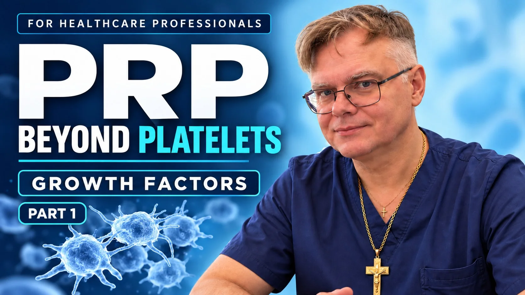
Watch the presentation
This first episode examines PDGF, TGF-β, VEGF, IGF-1, EGF and FGF-2. Continue with Part 2 — what is in the plasma?.
Open the full video on YouTube · English audio · Chapter links below
How an orthopaedic surgeon interprets PRP biology
PRP delivers a mixture of growth factors, cytokines and other bioactive proteins. Their functions overlap, and the response depends on concentration, receptors, the condition of the tissue and the timing of exposure. A platelet count describes one characteristic of a preparation; it is not a complete measure of its biological activity or expected clinical effect.
The network and release curves in the film are conceptual illustrations. They are not a universal timetable showing fixed peaks after every PRP injection. Preparation, activation, fibrin formation and local cells influence the availability of the signals.
Six growth factors, different contributions
PDGF — Cell recruitment and proliferation
Platelet-derived growth factor signals through PDGF receptors to influence chemotaxis, proliferation and extracellular matrix production. Its role extends across fibroblasts, stromal cells and vascular-supporting cells.
Clinical interpretation: Recruitment and collagen production are parts of repair. Fibre alignment, matrix maturation and appropriate mechanical loading still matter for functional tissue.
TGF-β — Matrix regulation and remodelling
Transforming growth factor beta regulates matrix synthesis, differentiation, immune activity and tissue remodelling. Its effects depend on cell type and the surrounding signals.
Clinical interpretation: Matrix deposition and restoration of native architecture are different outcomes. Excessive or persistent signalling can favour fibrosis; TGF-β should not be described simply as an anti-inflammatory factor.
VEGF — Vascular responses
Vascular endothelial growth factor influences endothelial migration, proliferation and survival, as well as vascular permeability. These mechanisms contribute to vascular responses during repair.
Clinical interpretation: More vascularisation is not universally beneficial. The meaning of a vascular response depends on the tissue, its condition and the timing and duration of the signal.
IGF-1 — Growth and cell survival
Insulin-like growth factor 1 supports anabolic and survival pathways in responsive cells. It also circulates in plasma, where IGF-binding proteins influence its availability.
Clinical interpretation: Support for cell metabolism or matrix synthesis does not itself demonstrate restoration of damaged cartilage. IGF-1 availability cannot be inferred from platelet concentration alone.
EGF — Cell migration and epithelial repair
Epidermal growth factor acts through EGFR to influence cell migration, proliferation and survival. Re-epithelialisation is particularly relevant to wound biology.
Clinical interpretation: Re-epithelialisation refers to an epithelial surface, such as epidermis. Synovium is not a conventional epithelial lining; experimental cellular effects do not establish clinical tendon regeneration.
FGF-2 — Proliferation, angiogenesis and matrix turnover
Fibroblast growth factor 2, also called basic fibroblast growth factor, influences proliferation, vascular responses and extracellular matrix turnover. Its activity intersects with other repair pathways.
Clinical interpretation: Increasing cell number does not guarantee a desired phenotype or a mechanically competent matrix. Receptor profiles, concentration and tissue context influence the response.
From mechanism to clinical interpretation
A mechanism provides a rationale for investigating a treatment. It does not, by itself, establish clinical effectiveness or prove structural regeneration. Responses observed in one tissue or experimental system cannot automatically be transferred to another clinical indication.
When interpreting a PRP study or preparation, consider the diagnosis and target tissue alongside platelet dose, plasma volume, leukocyte content, residual red cells, activation and handling. Delivery technique, rehabilitation and the selected clinical outcomes also belong in the assessment.
PRP is not a stem-cell concentrate and should not be presented as a guaranteed way to rebuild cartilage, tendon or bone. The useful question is whether a specified preparation produces a meaningful benefit for a specified indication.
Continue with Part 2: the plasma itself
Part 2 examines the bioactive environment beyond platelets: plasma proteins, cytokines, chemokines, fibrin-related components and their interactions. Part 2 is available. Read the Part 2 guide and watch the presentation.
Further reading
Platelet Rich Plasma: a short overview of certain bioactive components — background on PRP constituents and their biological functions.
Platelet-Rich Plasma: Where Are We Now and Where Are We Going? — biological rationale, preparation variability and the distinction between laboratory findings and clinical applications.
Related professional resources
Outpatient and Ultrasound-Guided Procedures · Musculoskeletal Ultrasound · Diagnostics and Clinical Pathways
Before considering PRP: turn the biology into a clinical question
A useful consultation starts with the diagnosis and the outcome that matters to the patient. “Improve the biology” is not a measurable treatment goal. Less pain on stairs, returning to a particular activity or reducing a specific limitation are clearer goals.
A worksheet for patients and clinicians
- Define the condition. Which tissue is being treated, how long has the problem been present and what supports the diagnosis?
- Describe care already tried. Record exercise, load modification, medicines or previous procedures, their duration and their effects.
- Identify the evidence. Ask whether the research concerns the same diagnosis and a comparable preparation. A study of knee osteoarthritis cannot automatically answer a question about a torn tendon.
- Specify the intended benefit. Separate pain and function from imaging change and structural regeneration. Ask which outcome will actually be measured.
- Agree on follow-up. What activity plan accompanies treatment, when will progress be reviewed and what happens if there is no meaningful improvement?
Questions about the proposed procedure
Ask about expected discomfort, risks, total cost, the number of injections being proposed and the alternatives. Discuss current medicines with the treating clinician; do not stop prescribed treatment independently to prepare for an injection. Request written instructions for aftercare and contact details for unexpected symptoms.
For clinicians, recording baseline function and the actual product used makes follow-up more interpretable than documenting only “PRP given”. For patients, the central question remains whether the expected benefit is worthwhile for their own problem.
Further reading: AAOS — PRP: indications, uncertainties and preparation differences.
Medical author: Tomasz Poboży - orthopaedic surgeon, MD, PhD — specialist in orthopaedics and musculoskeletal traumatology.
Professional educational material. It does not replace clinical judgement or individual patient assessment. Published: 9 October 2026.