 Skip to content
FOR CLINICIANS AND PHYSIOTHERAPISTS
Osteotomies around the knee — rehabilitation protocol
After osteotomy, restoration of motion and strength must be combined with protection of healing bone. The correction, fixation stability and follow-up findings determine loading, rather than the presence of a plate alone.
Using and loading the limb
HTO and DFO require different pathways. Early loading applies to selected stable HTOs; DFO often needs longer protection and healing review before progression.
Full guidance ↓Brace or immobilisation
Brace, lock and weaning depend on osteotomy type, fixation and additional repairs. Do not impose one schedule on every case.
Full guidance ↓Permitted movement
Set motion separately from loading. Meniscal, cartilage or additional stabilisation procedures may require their own restrictions.
Full guidance ↓Permitted movement, loading and removal of a brace are separate decisions. Additional repairs may require a more protective programme.
Rehabilitation at a glance
Progression follows healing and functional criteria, not dates alone.
Rehabilitation boards
Exercises — a closer look
Use the complete boards above together with the instructions for the chosen stage. An exercise shown for a later stage is not permitted early.
Open the relevant complete board and zoom in. The viewer retains all stages and captions.
5. How to perform the exercises
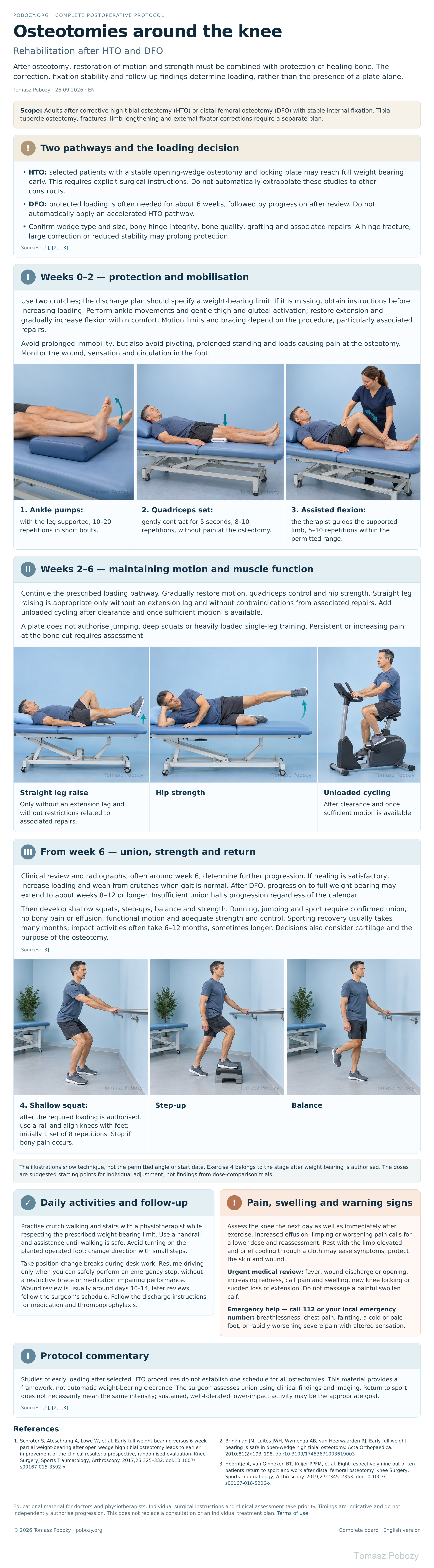
- 1. Ankle pumps: with the leg supported, 10–20 repetitions in short bouts.
- 2. Quadriceps set: gently contract for 5 seconds, 8–10 repetitions, without pain at the osteotomy.
- 3. Assisted flexion: the therapist guides the supported limb, 5–10 repetitions within the permitted range.
- 4. Shallow squat: after the required loading is authorised, use a rail and align knees with feet; initially 1 set of 8 repetitions. Stop if bony pain occurs.
The illustrations show technique, not the permitted angle or start date. Exercise 4 belongs to the stage after weight bearing is authorised. The doses are suggested starting points for individual adjustment, not findings from dose-comparison trials.
Scope and protection
1. Two pathways and the loading decision
- HTO: selected patients with a stable opening-wedge osteotomy and locking plate may reach full weight bearing early. This requires explicit surgical instructions. Do not automatically extrapolate these studies to other constructs.
- DFO: protected loading is often needed for about 6 weeks, followed by progression after review. Do not automatically apply an accelerated HTO pathway.
- Confirm wedge type and size, bony hinge integrity, bone quality, grafting and associated repairs. A hinge fracture, large correction or reduced stability may prolong protection.
Stages and progression criteria
2. Weeks 0–2 — protection and mobilisation
Use two crutches; the discharge plan should specify a weight-bearing limit. If it is missing, obtain instructions before increasing loading. Perform ankle movements and gentle thigh and gluteal activation; restore extension and gradually increase flexion within comfort. Motion limits and bracing depend on the procedure, particularly associated repairs.
Avoid prolonged immobility, but also avoid pivoting, prolonged standing and loads causing pain at the osteotomy. Monitor the wound, sensation and circulation in the foot.
3. Weeks 2–6 — maintaining motion and muscle function
Continue the prescribed loading pathway. Gradually restore motion, quadriceps control and hip strength. Straight leg raising is appropriate only without an extension lag and without contraindications from associated repairs. Add unloaded cycling after clearance and once sufficient motion is available.
A plate does not authorise jumping, deep squats or heavily loaded single-leg training. Persistent or increasing pain at the bone cut requires assessment.
4. From week 6 — union, strength and return
Clinical review and radiographs, often around week 6, determine further progression. If healing is satisfactory, increase loading and wean from crutches when gait is normal. After DFO, progression to full weight bearing may extend to about weeks 8–12 or longer. Insufficient union halts progression regardless of the calendar.
Then develop shallow squats, step-ups, balance and strength. Running, jumping and sport require confirmed union, no bony pain or effusion, functional motion and adequate strength and control. Sporting recovery usually takes many months; impact activities often take 6–12 months, sometimes longer. Decisions also consider cartilage and the purpose of the osteotomy.
Sources: [3]
Daily activities and follow-up
Progress in everyday activities follows the permitted loading and motion for your procedure. Walking without aids requires a controlled gait; returning to driving requires safe vehicle control and no impairing medication. The detailed recommendations below explain the criteria and exceptions.
6. Daily activities and follow-up
Practise crutch walking and stairs with a physiotherapist while respecting the prescribed weight-bearing limit. Use a handrail and assistance until walking is safe. Avoid turning on the planted operated foot; change direction with small steps.
Take position-change breaks during desk work. Resume driving only when you can safely perform an emergency stop, without a restrictive brace or medication impairing performance. Wound review is usually around days 10–14; later reviews follow the surgeon’s schedule. Follow the discharge instructions for medication and thromboprophylaxis.
Warning signs
7. Pain, swelling and warning signs
Assess the knee the next day as well as immediately after exercise. Increased effusion, limping or worsening pain calls for a lower dose and reassessment. Rest with the limb elevated and brief cooling through a cloth may ease symptoms; protect the skin and wound.
Urgent medical review: fever, wound discharge or opening, increasing redness, calf pain and swelling, new knee locking or sudden loss of extension. Do not massage a painful swollen calf.
Emergency help — call 112 or your local emergency number: breathlessness, chest pain, fainting, a cold or pale foot, or rapidly worsening severe pain with altered sensation.
References
References
- Schröter S, Ateschrang A, Löwe W, et al. Early full weight-bearing versus 6-week partial weight-bearing after open wedge high tibial osteotomy leads to earlier improvement of the clinical results: a prospective, randomised evaluation. Knee Surgery, Sports Traumatology, Arthroscopy. 2017;25:325–332. doi:10.1007/s00167-015-3592-x
- Brinkman JM, Luites JWH, Wymenga AB, van Heerwaarden RJ. Early full weight bearing is safe in open-wedge high tibial osteotomy. Acta Orthopaedica. 2010;81(2):193–198. doi:10.3109/17453671003619003
- Hoorntje A, van Ginneken BT, Kuijer PPFM, et al. Eight respectively nine out of ten patients return to sport and work after distal femoral osteotomy. Knee Surgery, Sports Traumatology, Arthroscopy. 2019;27:2345–2353. doi:10.1007/s00167-018-5206-x
Clinical commentary and terms of use
8. Protocol commentary
Studies of early loading after selected HTO procedures do not establish one schedule for all osteotomies. This material provides a framework, not automatic weight-bearing clearance. The orthopaedic surgeon assesses union using clinical findings and imaging. Return to sport does not necessarily mean the same intensity; sustained, well-tolerated lower-impact activity may be the appropriate goal.