 Skip to content
CONDITIONS AND SYMPTOMS
Tendons and Enthesopathies
Tendon disorders and enthesopathies may cause pain during walking, running, gripping, manual work or repeated loading. This page helps you find the relevant clinical problem. It does not replace detailed guides to individual tendons and attachments.
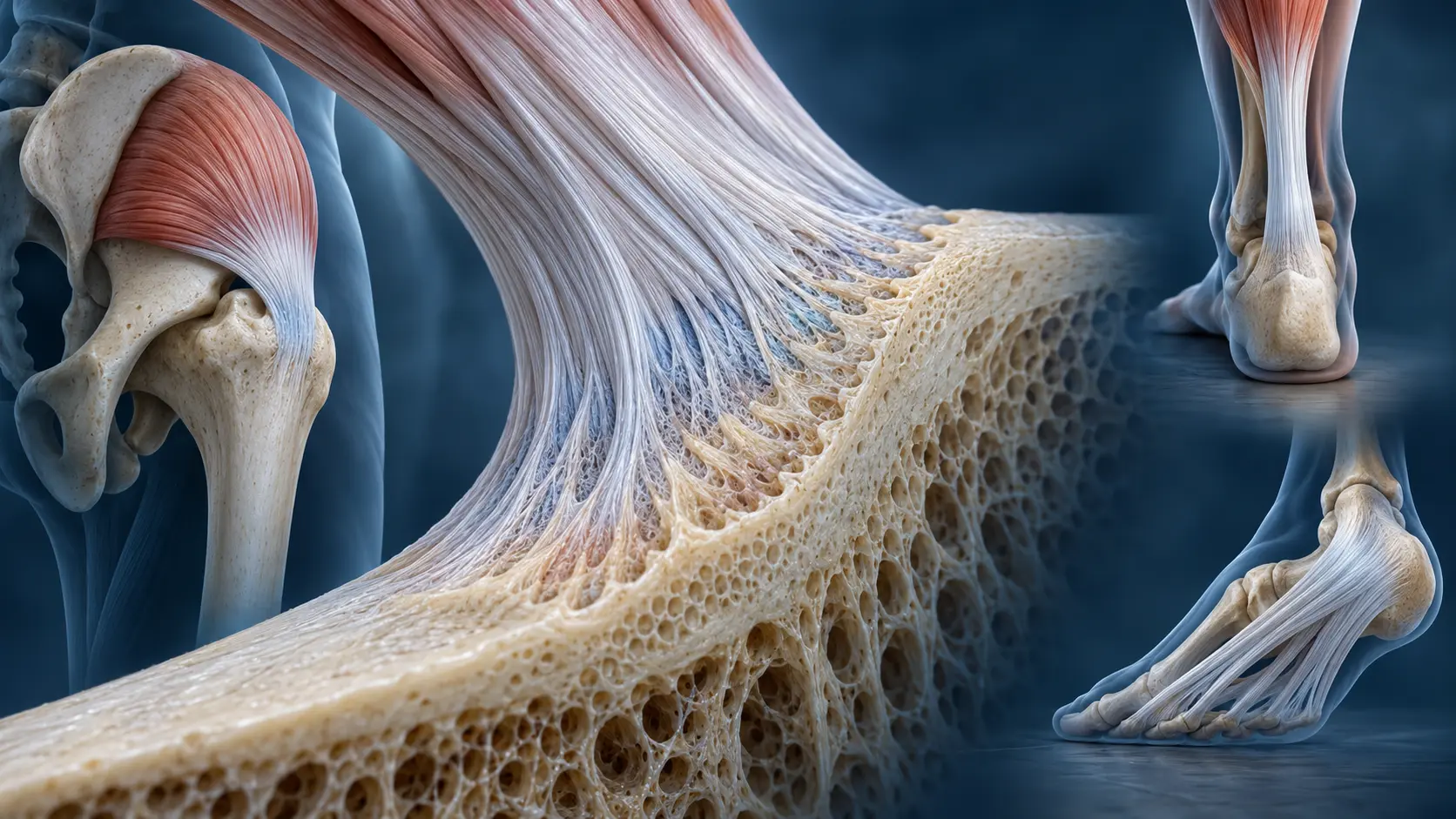
How do tendinopathy and enthesopathy differ?
Tendinopathy affects the tendon, while enthesopathy affects its attachment to bone. In practice, these areas overlap; terminology depends on the location and clinical picture. Not every pain near a tendon indicates tendon disease — similar symptoms may arise from a joint, bursa, nerve, bone or adjacent tissues.
Common problems
- Gluteal tendons and lateral hip pain (in Polish) — symptoms often worsen during side-lying, stair use or prolonged loading.
- Patellar tendon — “jumper’s knee” (in Polish) — pain is usually related to running, jumping and other dynamic loading.
- Semimembranosus tendon (in Polish) — one possible source of posteromedial knee pain.
- Achilles tendon (in Polish) — load-related pain and stiffness are typical; a sudden snap and marked loss of strength requires assessment for rupture.
- Posterior tibial tendon (in Polish) — pain and swelling on the inner side of the ankle may coexist with impaired arch function.
- Peroneal tendons (in Polish) — pain behind the lateral malleolus requires differentiation between tendinopathy, tearing and tendon instability.
- Plantar fascia (in Polish) — a common cause of heel pain, particularly during the first steps after rest.
- Tennis elbow (in Polish) and golfer’s elbow (in Polish) — overuse problems at tendon attachments around the elbow.
- Long head of biceps tendon (in Polish) — one possible source of anterior shoulder pain.
How does an orthopaedic surgeon assess a tendon problem?
Pain location, provoking activities, loading history and examination are key. Imaging is selected for a specific question. Ultrasound shows many superficial tendons and attachments well and allows selected structures to be examined dynamically. MRI may be needed for deeper abnormalities, suspected extensive injury or assessment of the joint and other tissues. X-rays may be important for bone, calcification, the attachment or accompanying joint changes.
Not every abnormal tendon appearance on ultrasound or MRI causes pain. Findings must be correlated with symptoms and examination.
What does treatment involve?
For many tendinopathies, treatment centres on appropriate load modification and gradual restoration of the tendon’s capacity to transmit force. Prolonged complete rest is not a universal solution. Exercise, load progression and return to activity depend on location, severity, symptom duration and patient demands.
Pain relief, braces, footwear modification, injections, shockwave therapy or surgery may be appropriate in selected situations, but no single programme suits all tendons. A procedure should be chosen for the diagnosis and treatment history, not solely an imaging report.
When may different investigations be needed?
Pain at tendon attachments may be related to overload, but can sometimes be part of inflammatory disease. If multiple attachments are painful, symptoms are bilateral or there is prolonged morning stiffness, joint swelling, psoriasis, uveitis, inflammatory bowel disease or systemic symptoms, assessment should not be limited to local tendon treatment.
A sudden injury with a snap, bruising, deformity or marked weakness may indicate an acute tear or rupture. These situations are discussed under Injuries and require separate assessment.
What did the study of 667 patients show?
In a study co-authored by Tomasz Poboży of 667 consecutive adults presenting with lower limb pain, ultrasound confirmed entheseal or tendon abnormalities in 22.8%. The gluteus medius tendon and plantar fascia were most often involved. These real-world outpatient data do not mean that ultrasound should automatically be performed for every painful limb, but illustrate the importance of tendon and attachment disorders in differential diagnosis.
Read the Polish discussion of the study.
References
- Poboży T, Janowski K, Domański P, et al. The Hidden Burden of Entheseal Pathology in Lower-Limb Pain: A Real-World Ultrasound Study of 667 Adults. Mayo Clin Proc Innov Qual Outcomes. 2026;10(4):100734. DOI: 10.1016/j.mayocpiqo.2026.100734.
This educational material does not replace individual examination or diagnosis.
Pain below the kneecap: plan the next step
Patellar tendinopathy commonly produces pain related to loading the tendon, particularly jumping and similar demanding activities. Pain in the same region can have other causes, so point to the painful area and explain which movements provoke it rather than assuming every symptom is “jumper’s knee”.
Prepare a loading history
Write down recent changes in training volume, intensity, surfaces, recovery and work demands. Include activities outside sport: stairs, lifting, kneeling and prolonged periods on your feet. Record what happens during activity, afterward and the next morning.
Ask for a graded programme
- Which activities should be reduced temporarily, and which can continue?
- What starting exercises and resistance suit my present capacity?
- What response is acceptable, and what means the load should be reduced?
- How will strength and function be reassessed?
- Which criteria should I meet before progressing to running, jumping or competition?
A programme should explain progression as well as the starting exercise. Complete rest may reduce symptoms without rebuilding capacity, while repeatedly pushing through worsening symptoms can make the plan unsustainable. Discuss changes with your physiotherapist instead of copying a high-load programme from an athlete at a different stage.
Imaging and procedures
Ask what ultrasound or MRI is expected to clarify and whether the result will change treatment. An abnormal tendon image does not independently determine how much pain you should have or when you can return to sport. If an injection or other procedure is proposed, ask what it adds to a loading programme and how benefit will be judged.
A sudden snap with bruising, marked weakness or inability to actively straighten the knee needs urgent assessment for a possible tear rather than routine tendinopathy exercises. See the full Polish guide to patellar tendon pain for further background.
Medical author
Tomasz Poboży - orthopaedic surgeon, MD, PhD — specialist in orthopaedics and musculoskeletal traumatology.
This educational material does not replace an individual consultation or medical examination. Last updated: 22 October 2026.