 Skip to content
FOR CLINICIANS AND PHYSIOTHERAPISTS
Arthroscopic subacromial decompression — rehabilitation protocol
Protection, movement and a gradual return to daily activity. Select the stage appropriate to the actual procedure and current clearance.
Using and loading the limb
Isolated decompression allows early light arm use. Progress resistance according to tolerance; this pathway does not cover tendon repair or tenodesis.
Full guidance ↓Brace or immobilisation
Use a sling briefly for comfort, without unnecessary prolonged immobilisation. Protect the numb limb while the nerve block remains active.
Full guidance ↓Permitted movement
Passive, assisted and active movement follow control and comfort. Isolated surgery does not routinely require waiting several weeks for active motion.
Full guidance ↓Permitted movement, loading and removal of a brace are separate decisions. Additional repairs may require a more protective programme.
Rehabilitation at a glance
Progression follows healing and functional criteria, not dates alone.
Rehabilitation boards
This pathway applies to isolated decompression. Tendon repair, tenodesis, labral repair or another reconstruction adds its own protection requirements from the start.
Early movement and range of motion
From day one · priorities: mobility and comfort
1Brief sling use
Usually 1–3 days for comfort. Also protect the arm until the nerve block resolves. Wean as early as comfortable; persistent dependence needs review. [1–3]
2Start moving early
Once sensation and control return: assisted, then active movement as tolerated. No routine 90° limit. Do not force movement through severe pain. [3,6]
3First 0–2 weeks
Short daily sessions; progressively regain elevation and rotation. Use the arm for light daily tasks. Avoid heavy lifting and overload.
4Physiotherapy in week one
Instruction before discharge, followed by assessment, movement assistance and technique correction. Home exercise complements treatment. [2]
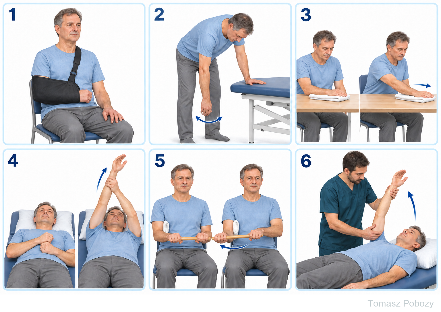
© 2026 Tomasz Poboży – orthopaedic surgeon · pobozy.org
Full movement, strength and activity
Approximately weeks 2–12 and beyond · progress according to function
1Around weeks 2–6
Work towards full passive and active movement without shoulder hiking. If movement returns earlier, use it rather than waiting for a calendar milestone.
2Individualised strengthening
Gentle pain-free isometrics may start early. Introduce low resistance when control and tolerance allow, often during weeks 2–6. [5]
3From around week 6
Develop strength, endurance and work-specific tasks. Heavy or repetitive overhead loading often requires 8–12 weeks or longer. [2,6]
4Return criteria
Functionally full comfortable movement, scapular control, task-appropriate strength and no clear deterioration after a trial or the following day.
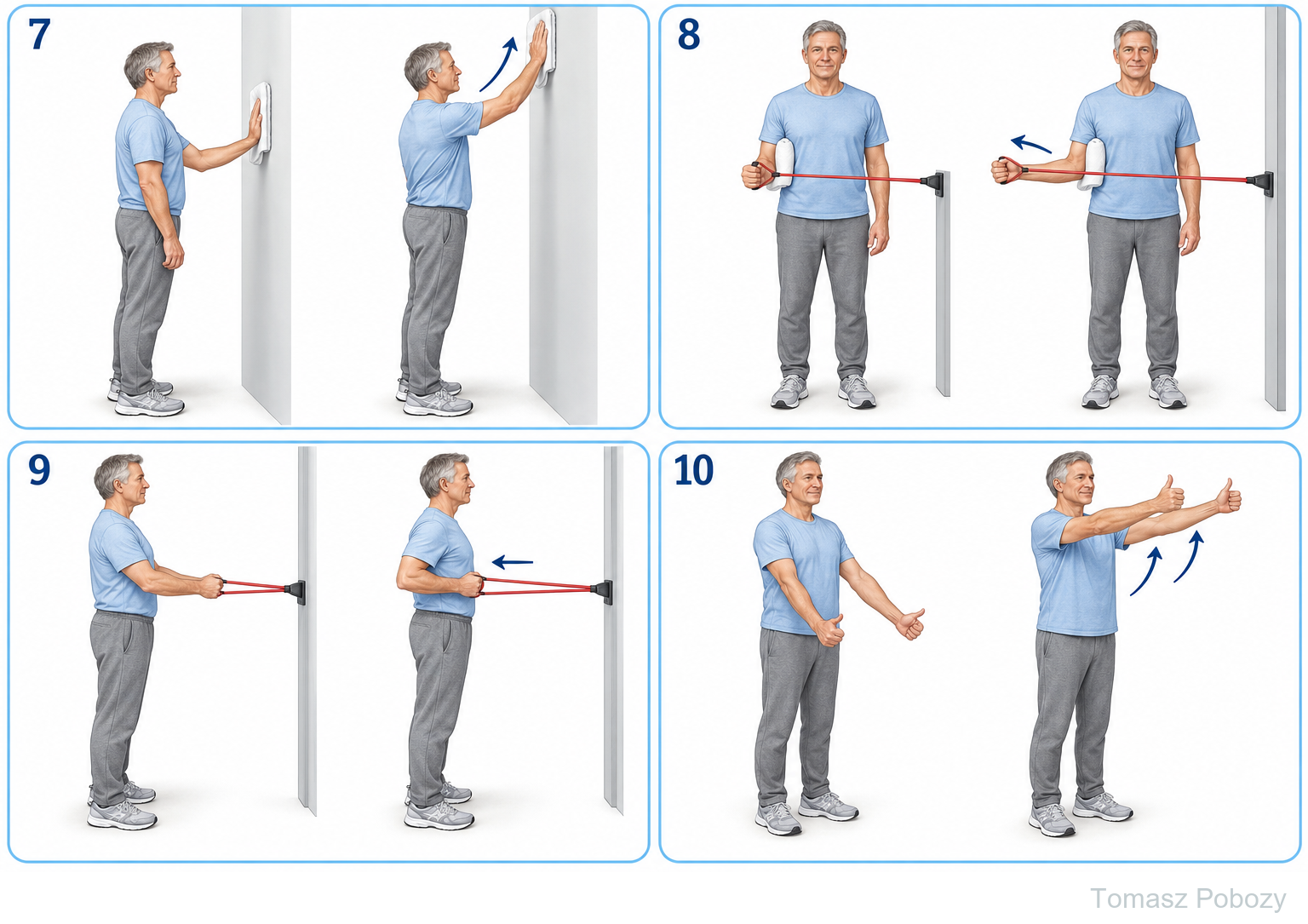
© 2026 Tomasz Poboży – orthopaedic surgeon · pobozy.org
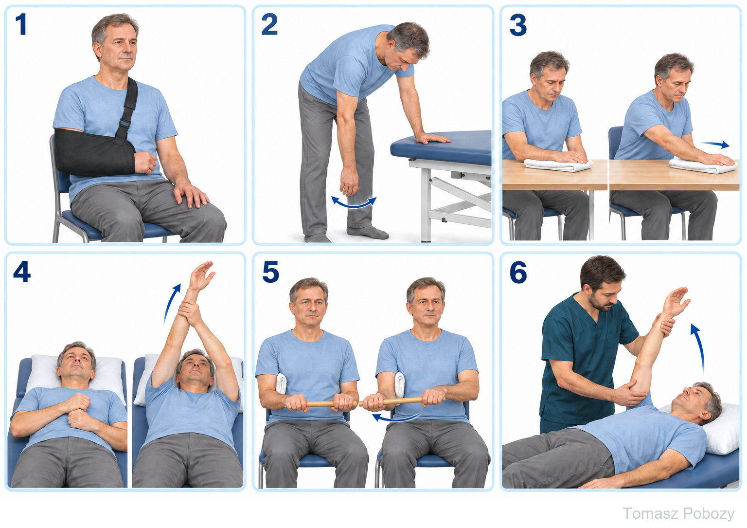
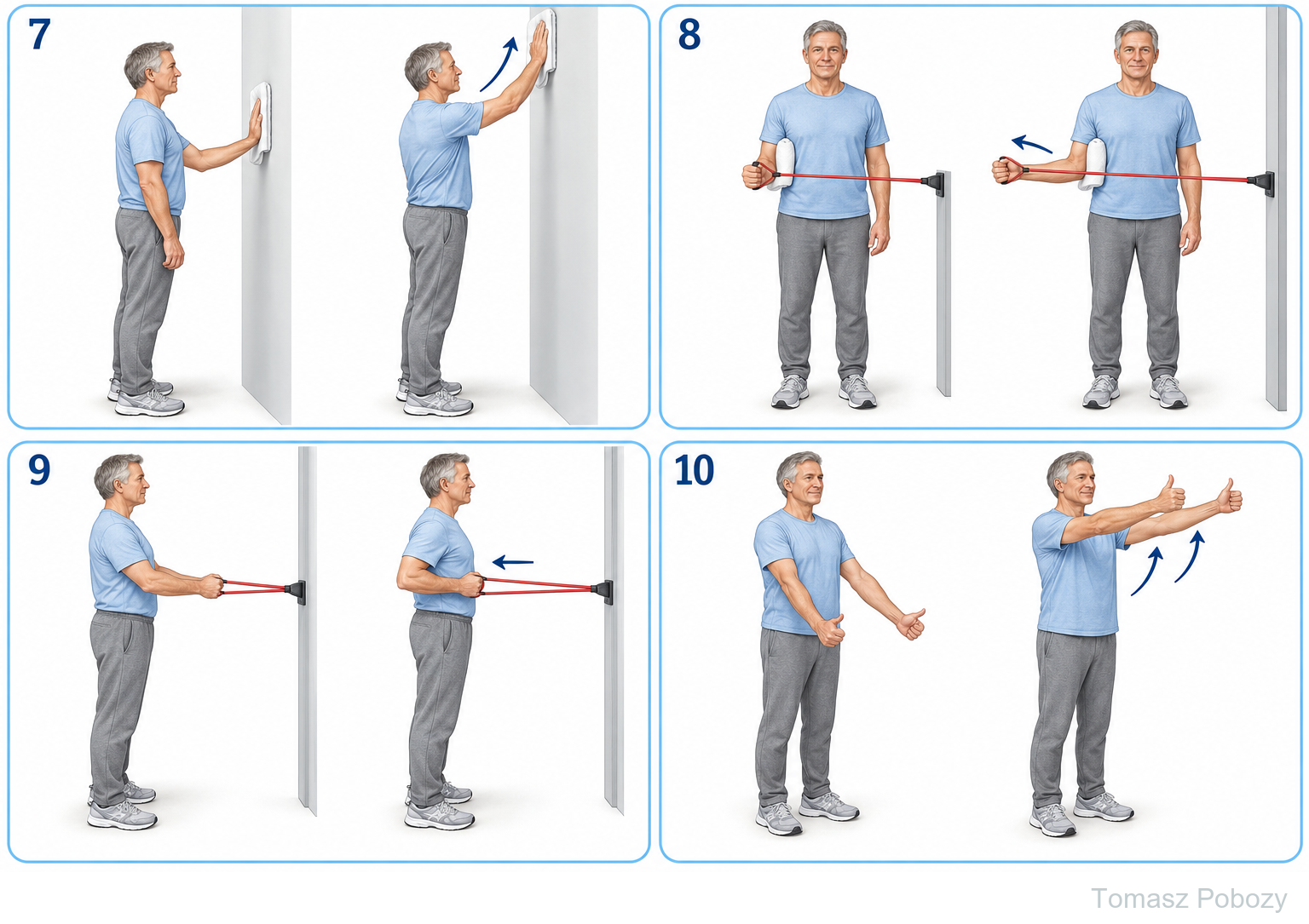
Exercises — a closer look
Use the complete boards above together with the instructions for the chosen stage. An exercise shown for a later stage is not permitted early.
Exercise technique and symptom response
The physiotherapist selects passive, assisted and active exercises. Progression to active movement depends on control and comfort, without a mandatory wait of several weeks. Add elbow, wrist and finger movement and gentle scapular mobility. [5,6]
Mild stretching discomfort may be acceptable. Sharp pain, marked guarding or a clear increase in symptoms lasting more than about 30 minutes calls for less range, fewer repetitions or lower resistance. Also review sleep and next-day symptoms. Do not force end range. [3,4]
Scope and protection
Sling, pain and wound care
The sling provides comfort rather than weeks of immobilisation. Protect the numb arm while the block is active. Start independent shoulder exercises when control returns; earlier movement requires instructions from the clinical team.
Take medication as prescribed. A cold pack through fabric may help; do not apply to numb skin or wet the dressing. A pillow under the forearm may improve sleep. Follow the orthopaedic surgeon’s wound-care instructions. [1,2,5]
The physiotherapist’s role and progression
At the initial visit assess the wound, pain, passive and active range, scapulohumeral rhythm and independence. Assist movement, choose supported positions, check home exercises and document progress. Visit frequency depends on need; treatment is not limited to passive modalities.
Aim to regain full movement as early as tolerated, without a universal deadline. Lack of progress or increasing stiffness in the first 2–4 weeks warrants reassessment. Before increasing resistance, check movement quality and response to the previous dose.
Daily activities and follow-up
Daily activities follow the permitted shoulder and elbow motion, lifting and arm support for your procedure. Sling removal is not clearance for lifting or driving. The detailed guidance below explains the criteria and exceptions.
Work, driving and sport
Desk work is often possible within 1–3 weeks depending on symptoms and access to breaks. Manual work usually takes several weeks to a few months; heavy overhead work may require 3 months or longer. These are guides, not automatic clearance. [2,5]
Resume driving only without a sling or impairing medication, with safe steering control and the ability to perform an emergency manoeuvre. Start sport with brief trials, then increase volume and intensity. Comfortable elevation does not establish readiness for throwing, heavy lifting or racket sports.
Warning signs
When should exercise stop?
Urgent medical review: fever, wound drainage, increasing redness, rapidly worsening pain or new weakness or numbness persisting beyond the expected duration of the block. Do not exercise through these symptoms. [4]
Emergency assistance: breathlessness, chest pain, collapse or a cold, pale hand. Stop treatment and seek emergency help.
For a life-threatening emergency, call your local emergency number immediately — 112 in the European Union. On the boards, 112 refers to the EU.
Additional scope notes
Rehabilitation protocol
Brief sling use. Early movement. Physiotherapist-guided exercise and a gradual return to function.
Watch or listen · Polish recordings
Educational recordings linked by Tomasz Poboży — orthopaedic surgeon in the Polish version: diagnosis and treatment options, not an individual postoperative prescription.
Shoulder pain and rotator cuff tears — Tomasz Poboży — orthopaedic surgeon · YouTube · PL
References
References
- Royal Orthopaedic Hospital. Subacromial Decompression (SAD).
- Hull University Teaching Hospitals NHS Trust. Arthroscopic Subacromial Decompression. 2025.
- North Tees and Hartlepool NHS Foundation Trust. Subacromial Decompression and/or Excision of the Distal Clavicle. Stage 1 — Physiotherapy Advice and Exercise.
- University Hospital Southampton NHS Foundation Trust. Arthroscopic subacromial decompression: exercises.
- North Tees and Hartlepool NHS Foundation Trust. Arthroscopic Sub acromial Decompression +/- ACJ excision. 2023.
- Worcestershire Acute Hospitals NHS Trust. Advice and exercises following Subacromial decompression.
Clinical commentary and terms of use
Commentary
Following isolated decompression, the priority is to restore full movement as early as tolerated. Brief sling use can help manage pain but should not delay mobilisation.
Working with a physiotherapist provides exercise assistance and appropriate progression. The patient continues the learned programme at home. The surgeon communicates the operative findings, and the physiotherapist reports progress and difficulties.
The stages are a practical synthesis of institutional guidance, which differs particularly on return to loading. They are not a single validated schedule for every patient. Early full movement and progressive strengthening have different progression criteria.
Use of this protocol
Educational material for doctors and physiotherapists. Requires patient assessment, the operative report and individual instructions. Does not replace a treatment plan or ongoing monitoring. Illustrations are examples, not the required range at the first session.
© 2026 Tomasz Poboży – orthopaedic surgeon · pobozy.org