 Skip to content
FOR CLINICIANS AND PHYSIOTHERAPISTS
Rotator cuff repair — rehabilitation protocol for small and medium tears
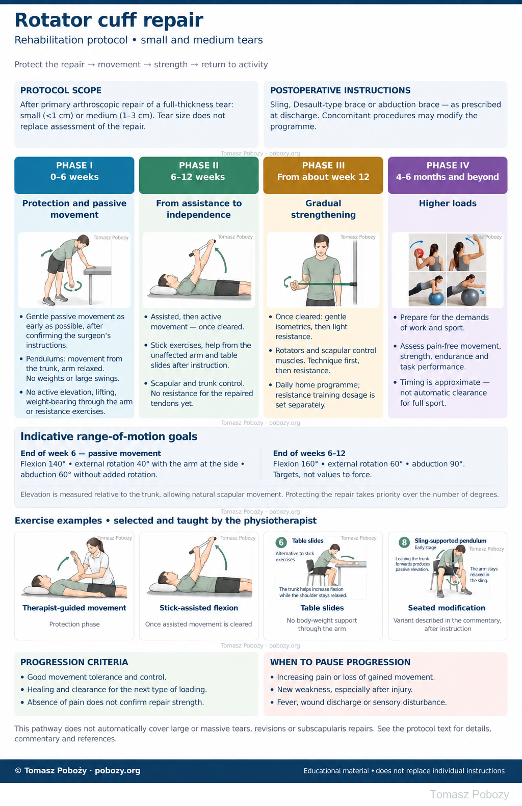
After rotator cuff repair, rehabilitation protects the repair site and gradually restores shoulder movement and function. The pathway below uses early passive movement for small and medium tears. The orthopaedic surgeon’s instructions in the discharge summary take priority over the general programme.1, 2
Using and loading the limb
Weeks 0–6: no active arm elevation, lifting, body-weight support or resistance for the repaired tendons. Strengthening requires clearance, around week 12 in this pathway.
Full guidance ↓Brace or immobilisation
Sling, Desault support or abduction brace: type and duration depend on the repair and discharge instructions. Less pain alone does not permit discontinuation.
Full guidance ↓Permitted movement
Begin with passive movement. End-of-week-6 targets in this pathway: elevation 140°, ER 40°, abduction 60°; progress gently and respect any stricter prescription.
Full guidance ↓Permitted movement, loading and removal of a brace are separate decisions. Additional repairs may require a more protective programme.
Rehabilitation at a glance
Progression follows healing and functional criteria, not dates alone.
Rehabilitation boards
Exercises — a closer look
Use the complete boards above together with the instructions for the chosen stage. An exercise shown for a later stage is not permitted early.
Open the relevant complete board and zoom in. The viewer retains all stages and captions.
Scope and protection
Scope, immobilisation and postoperative instructions
After primary arthroscopic repair of a full-thickness rotator cuff tear classified as small or medium. In a size-based classification, a small tear measures less than 1 cm and a medium tear 1–3 cm. This is the size of the tear, not the width of a normal tendon. The documentation should specify how it was measured and which tendons were torn and repaired.5
This pathway does not automatically cover large or massive tears, extensive multi-tendon repairs, revision surgery or subscapularis repair. Concomitant procedures, such as biceps tenodesis, may alter the exercises permitted.
Start with the discharge summary
Immobilisation. Depending on tear size and repair conditions, the surgeon may prescribe a sling, a Desault-type immobiliser or an abduction orthosis. Its type, position and duration of use must follow the postoperative instructions.
Movement and loading. The permitted passive range and the timing of active arm elevation and resistance exercises must be known. Unclear or conflicting instructions require clarification with the surgeon.2
Pain control from the first days
During the first few days, baseline analgesics prescribed for regular use are taken at the scheduled times, as directed in the discharge summary, rather than waiting until pain becomes severe. This is particularly important as a nerve block wears off. Effective pain control supports rest and permitted exercises.3
Medication prescribed as needed remains as needed, particularly opioids. Treatment selection takes contraindications into account. The dose must not be increased to force a greater range of movement. Persistent severe pain requires reassessment.3
Stages and progression criteria
Phase I · weeks 0–6 — protection and passive movement
Protection and early passive movement
Gentle passive exercises begin as early as permitted,1 usually within the first week in this pathway, once the surgeon’s instructions have been confirmed. A longer period of protection prescribed for an individual repair remains binding.
The programme includes permitted hand, wrist and elbow movement, protected passive arm elevation and external rotation, and gentle Codman pendulum exercises. Gentle scapular positioning and control are also taught without loading the repair.
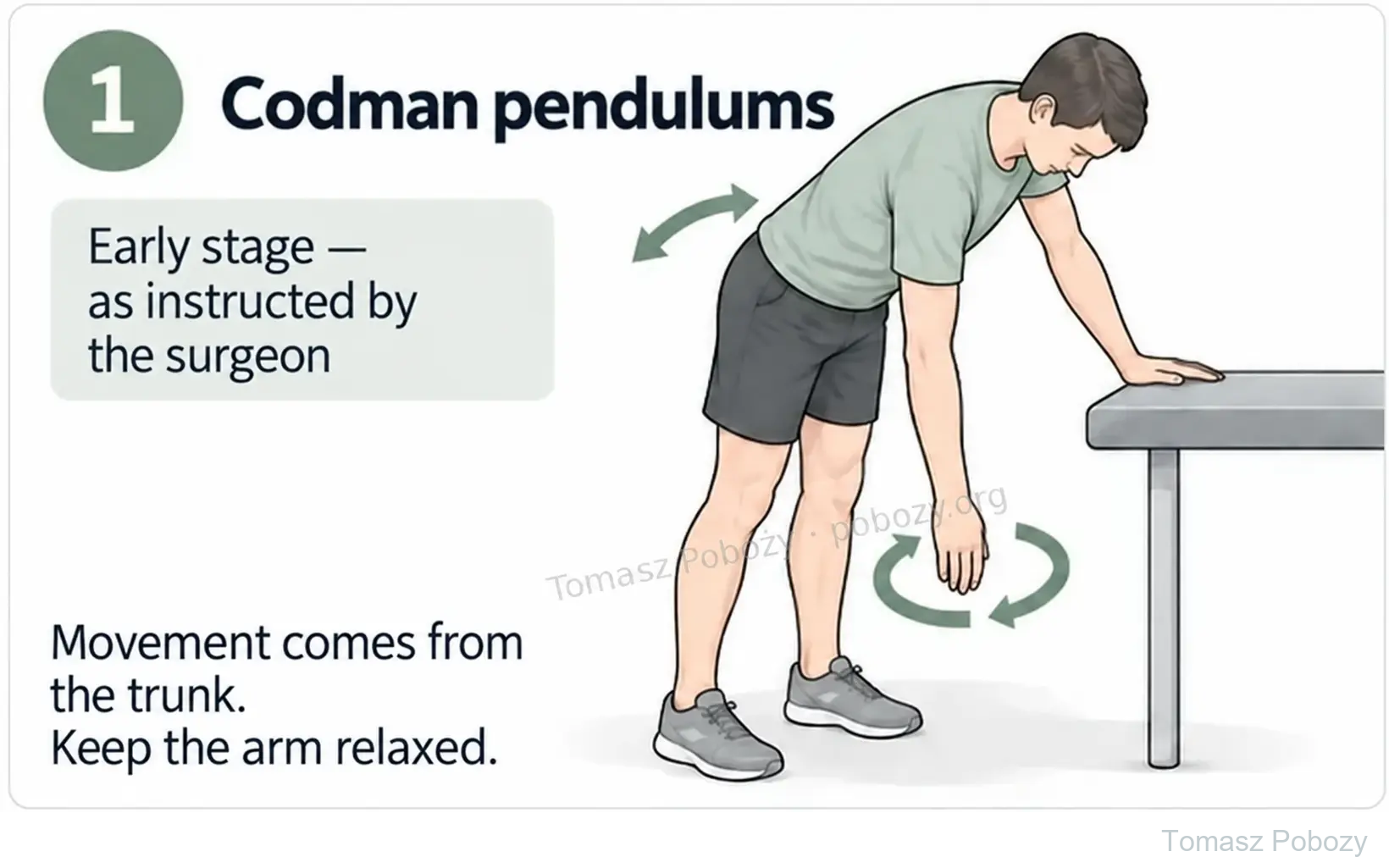
Codman pendulum exercises
The patient leans forward and supports the body with the unaffected hand on a stable surface. Small shifts in body weight gently move the freely hanging arm. No weight, large swings or active shoulder elevation are used.9
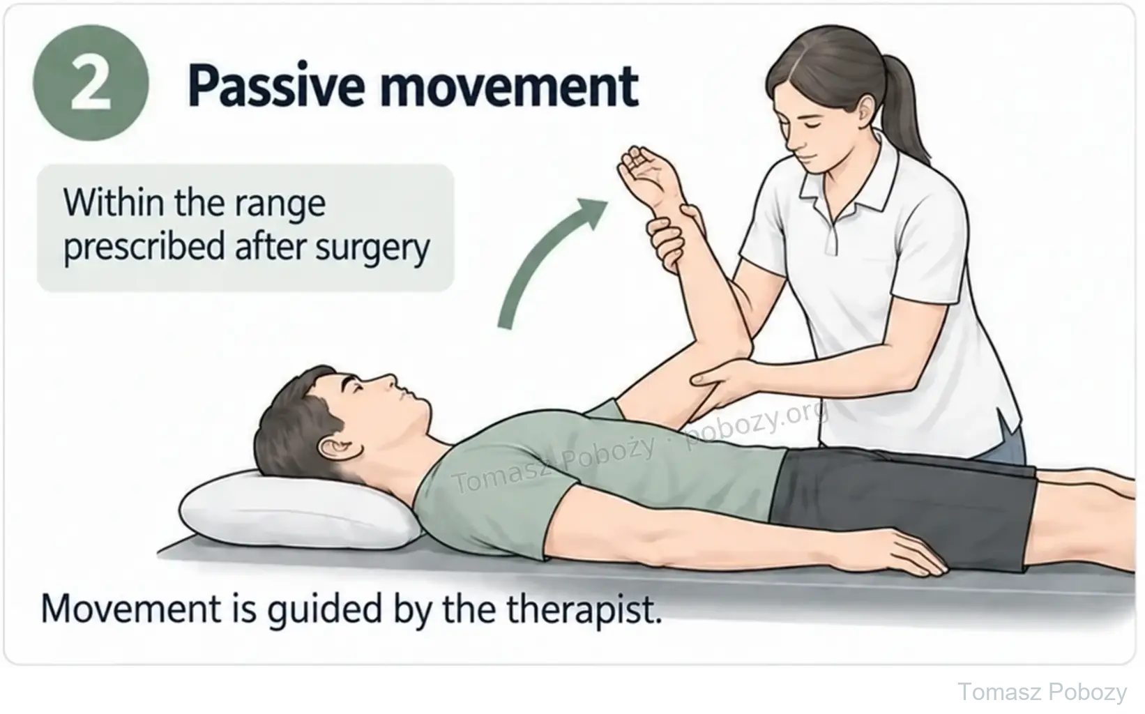
Passive movement with a physiotherapist
The patient relaxes the operated limb. The physiotherapist selects the hold and position and monitors the response to movement. Pendulums supplement rather than replace the full passive movement programme.9
Author-selected indicative range-of-motion targets
140° — flexion: raising the arm forwards.
40° — external rotation with the arm at the side.
60° — abduction without added rotation.
All three values are passive range-of-motion targets to work towards gradually by the end of week 6 in the author-selected pathway for medium tears. They are not first-session targets and must not be forced. The surgeon’s instructions and exercise tolerance may require slower progression.
Here, 140° of flexion means overall forward elevation of the arm relative to the trunk — 50° above shoulder level. Movement involves the glenohumeral joint and the other components of the shoulder complex, with natural scapular contribution. It is not rigidly divided into 90° at one joint and 50° elsewhere, nor achieved by leaning the trunk backwards.4
During this phase, avoid independently raising the operated arm, lifting objects, supporting body weight through that hand, and resistance exercises for the repaired tendons.10
Progression: around week 6, provided movement is well tolerated, there are no concerning symptoms and clinical assessment supports progression. Range must not be forced to meet a date in a table.10
Phase II · weeks 6–12 — from assisted to active movement
From assisted to independent movement
The aim is smooth movement without abruptly shrugging the shoulder towards the ear or substituting trunk leaning for arm movement. Exercises begin in a supported position, such as lying down, then progress to sitting and standing. The orthosis is discontinued according to the surgeon’s instructions.
The author-selected indicative targets by the end of this phase are flexion up to 160°, external rotation up to 60° and abduction up to 90°, with continued recovery towards full range. Resistance exercises for the repaired tendons are not yet introduced in this programme.
How should assisted exercises be performed?
The unaffected hand helps guide the operated arm without jerking or forcing against resistance. The physiotherapist demonstrates the exercise, selects the position and range and checks that the patient can reproduce it. After this preparation, the patient can perform the selected exercises independently at home. Not every example below needs to be performed.
Assisted exercises are introduced only once this stage has been cleared. A stick or the other hand is not a reason to advance the programme independently.
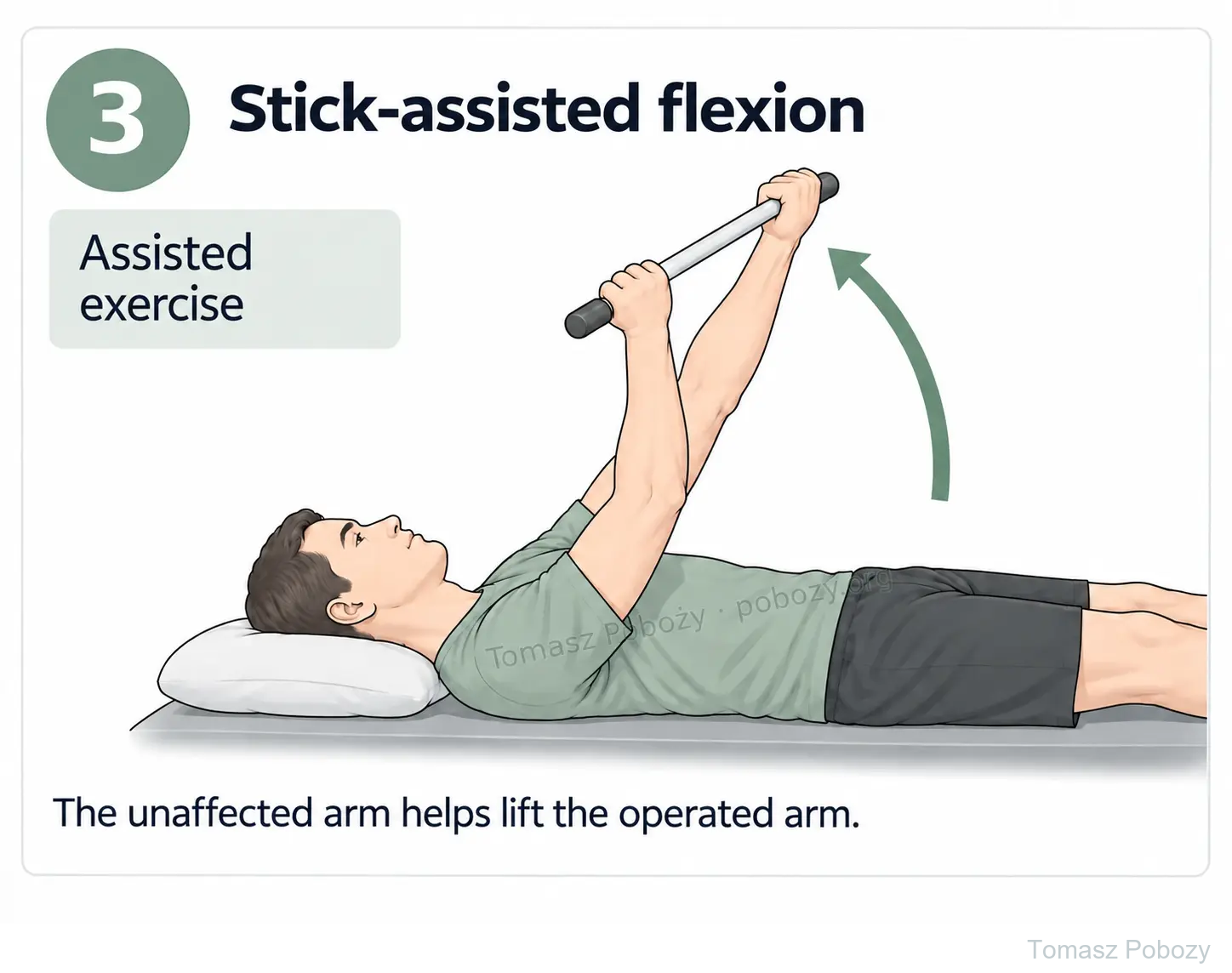
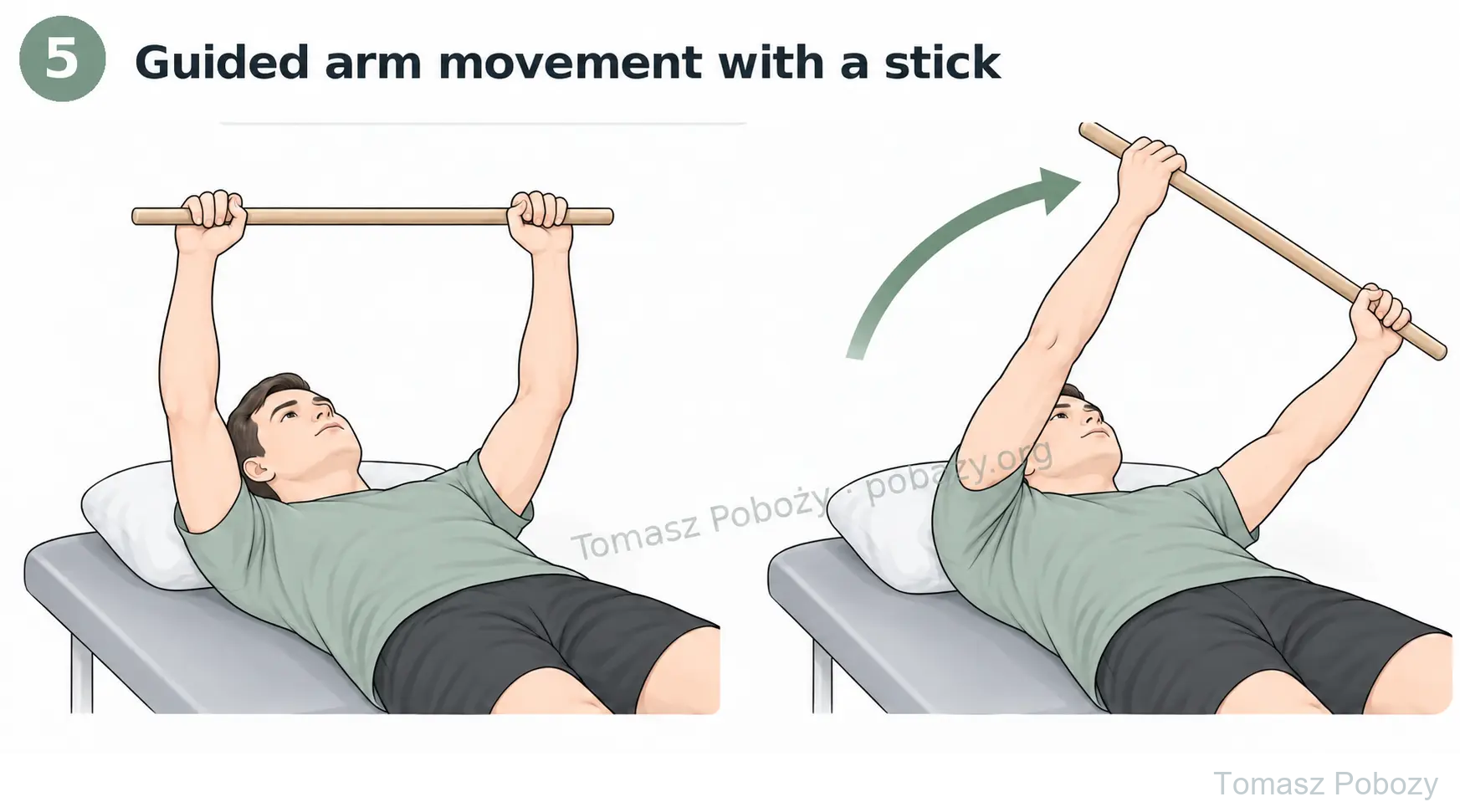
Stick-assisted flexion
Lying on the back, the patient holds a light stick with both hands. The unaffected arm helps move the stick upwards and return to the starting position. The range and elbow position are selected so that movement remains gentle and respects the repair restrictions.
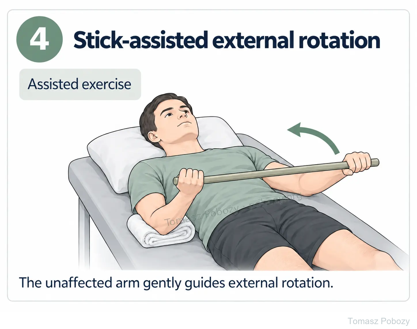
Stick-assisted external rotation
The patient lies on the back. The operated elbow is bent to approximately 90°, supported and kept close to the trunk. The unaffected hand uses the stick to guide the forearm outwards. The elbow must not move away from the body, the trunk must not rotate and the prescribed rotation limit must not be exceeded.
Guiding the arm in different directions
A stick may assist not only forward elevation but also movement in other selected directions, including abduction. Each movement must be taught separately; simply moving the stick from side to side is not a substitute. Large abduction and rotation must not be combined unless that movement has been cleared.
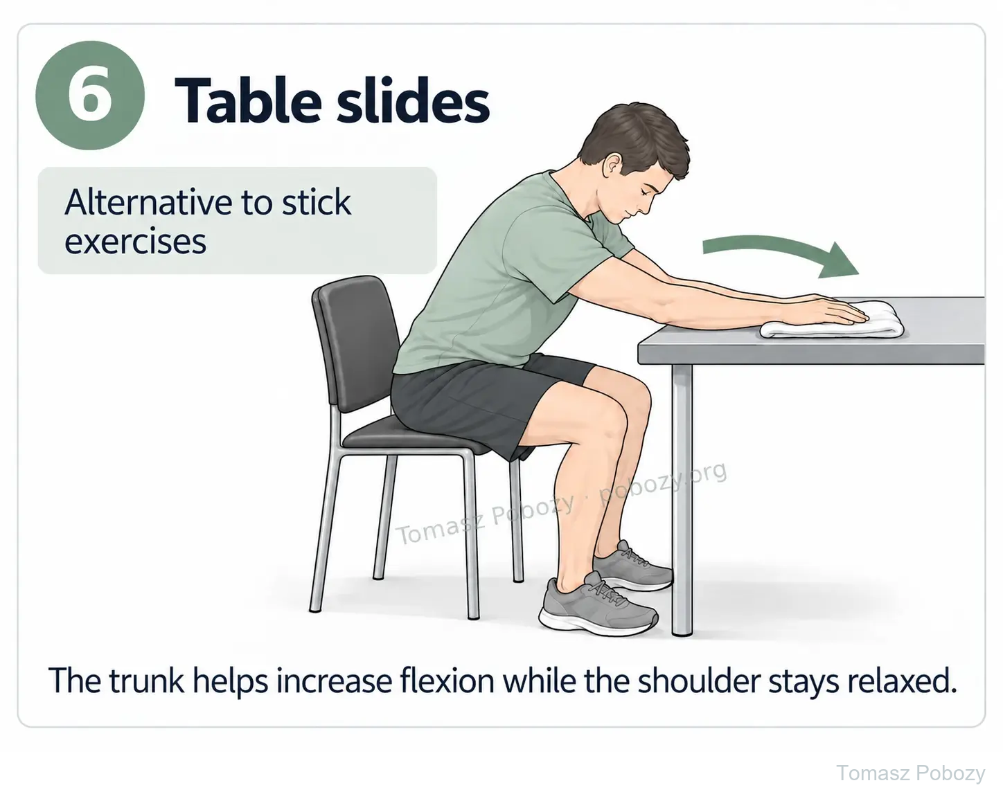
Table slides
The patient sits at a table with the limb supported on a towel. The unaffected hand assists while a controlled forward lean helps slide the hand forwards. Body weight must not be placed through the operated hand, and the arm must not be pushed through pain.
Assistance from the other hand — without a stick
Lying on the back, the patient supports the operated arm with the unaffected hand at the point shown by the physiotherapist. This assists elevation and a controlled return. Support under the elbow may be used when guiding a straighter arm is too difficult.
A rope-and-pulley device may be another aid after instruction and clearance for this stage. It is neither essential nor inherently gentler than a stick exercise.
Home programme: selected exercises in short sessions, with dosage and range agreed with the physiotherapist. The patient should know when to stop an exercise and what to report to the team. Increasing pain, clear deterioration in movement or persistently reduced tolerance calls for programme adjustment.
Progression to strengthening: around week 12, when active movement is pain-free, arm and scapular control are good and clinical assessment permits loading. Absence of pain alone does not replace the time needed for healing.10
Phase III · from approximately week 12 — gradual strengthening
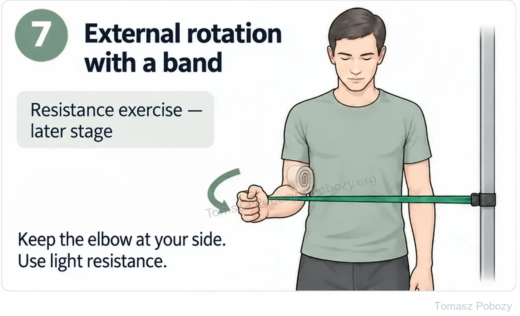
Once loading is permitted, gentle isometric exercises are introduced, followed by movement against light resistance. These involve the shoulder rotators and scapular control muscles. Correct movement comes before greater resistance.2, 10
At this stage, we combine physiotherapist-led sessions, for example three times a week, with a daily home programme. The physiotherapist adjusts appointment frequency, exercise selection, sets, repetitions and resistance to functional assessment and postoperative instructions. At home, the patient performs an individually prescribed daily set of mobility and arm-control exercises. Daily rehabilitation does not mean daily resistance training of the same muscles: its frequency and recovery intervals are set separately. Resistance is not increased if technique deteriorates or symptoms rise.
The elbow remains close to the trunk, with resistance low enough to allow movement without compensation. Bands are not used to stretch a fresh repair early in recovery.
Phase IV · around 4–6 months and beyond — higher loads
Tasks required for work and sport are introduced. Readiness depends not only on time since surgery but also on pain-free movement, strength, endurance and performance of the specific task.2, 10
The author-selected indicative criteria used here include at least 85% of the opposite side’s external- and internal-rotation strength. This is one criterion, not independent clearance for unrestricted sport. If the other shoulder is impaired, symmetry alone is also insufficient to assess function. Heavy work and overhead activities require gradual preparation and a decision by the treating team.
The 4–6-month period describes a stage of gradual return. It is not a guaranteed date for full recovery or clearance for unrestricted loading.
Daily activities
Support the forearm during rest, sleep and hygiene; follow the prescribed sling schedule. Dress the operated arm first and undress it last. Do not push out of a chair through the operated hand during the protection period. Desk work requires a supported arm, breaks and compliance with motion limits. Driving requires removal of the sling, permitted active control sufficient for emergency manoeuvres and no impairing medication. Heavy work and sport require procedure-specific progression and assessment.
Warning signs
Daily activity and warning signs
Starting active movement does not yet permit lifting, pushing, pulling or overhead work. Light desk work may be possible earlier if the arm is supported and restrictions are respected. Driving while immobilised should be avoided; discontinuing the orthosis alone does not establish readiness to drive safely.
When should progression stop?
Loading is not increased when pain rises, previously gained movement is lost or new weakness develops, particularly after an injury. Fever, wound discharge, persistent altered sensation and uncontrolled pain require contact with a doctor. A new phase is not started solely because the date in the table has arrived.
For a life-threatening emergency, call your local emergency number immediately — 112 in the European Union. On the boards, 112 refers to the EU.
Additional scope notes
Protecting the repair takes priority over the calendar. Timings and exercises apply to the group of repairs described. Using the programme requires knowledge of the operation and postoperative instructions, together with ongoing patient assessment. The board is a summary, not a substitute for the detailed description.
Passive movement takes place without intentional contraction of the operated shoulder muscles: the limb is moved by a therapist, a trained person or an appropriate aid. During active-assisted movement the patient contributes to moving the operated limb while receiving assistance. Active movement is performed independently. Using a stick or the other hand does not in itself make a movement fully passive.
Watch or listen · Polish recording
Tomasz Poboży — orthopaedic surgeon — Ból barku i uszkodzenie stożka rotatorów – od diagnozy do sprawnej ręki (PL)Background on shoulder pain, diagnosis and treatment. The recording does not replace procedure-specific postoperative restrictions.
References
References and scientific basis
Studies compare early and delayed mobilisation after arthroscopic rotator cuff repair. The pathway presented is one option, not the only valid timetable.1
The 140°/40°/60° ranges are author-selected indicative targets for the group of repairs described. The 2016 ASSET consensus describes another pathway with a longer initial period of protection.10
This is a synthesis based on the listed sources, not a separately validated programme from one centre. Research summaries do not replace assessment of an individual repair. The 2026 study by Kuş et al. suggests that selected low-supraspinatus-activity exercises may reduce early pain; however, the available abstract does not allow reconstruction of the full programme or assessment of structural safety. It does not justify arbitrary acceleration of rehabilitation.11
- Mazuquin B, Moffatt M, Gill P et al. Effectiveness of early versus delayed rehabilitation following rotator cuff repair: Systematic review and meta-analyses. PLoS One. 2021;16(5):e0252137. doi:10.1371/journal.pone.0252137.
- Sgroi TA, Cilenti M. Rotator cuff repair: post-operative rehabilitation concepts. Curr Rev Musculoskelet Med. 2018;11(1):86–91. doi:10.1007/s12178-018-9462-7.
- Toma O et al. PROSPECT guideline for rotator cuff repair surgery: systematic review and procedure-specific postoperative pain management recommendations. Anaesthesia. 2019;74:1320–1331. doi:10.1111/anae.14796.
- Braman JP et al. In vivo assessment of scapulohumeral rhythm during unconstrained overhead reaching in asymptomatic subjects. J Shoulder Elbow Surg. 2009;18:960–967. doi:10.1016/j.jse.2009.02.001.
- Tanaka M et al. Location and thickness of delaminated rotator cuff tears: cross-sectional analysis with surgery record review. JSES Open Access. 2018;2:84–90. doi:10.1016/j.jses.2017.11.004.
- Mochizuki T et al. Humeral insertion of the supraspinatus and infraspinatus. New anatomical findings regarding the footprint of the rotator cuff. J Bone Joint Surg Am. 2008;90:962–969. doi:10.2106/JBJS.G.00427.
- Lumsdaine W et al. Morphology of the humeral insertion of the supraspinatus and infraspinatus tendons: Application to rotator cuff repair. Clin Anat. 2015;28:767–773. doi:10.1002/ca.22548.
- Meyer DC et al. Quantitative analysis of muscle and tendon retraction in chronic rotator cuff tears. Am J Sports Med. 2012;40:606–610. doi:10.1177/0363546511429778.
- Cunningham G et al. Shoulder Motion Analysis During Codman Pendulum Exercises. Arthrosc Sports Med Rehabil. 2020;2:e333–e339. doi:10.1016/j.asmr.2020.04.013.
- Thigpen CA et al. The American Society of Shoulder and Elbow Therapists’ consensus statement on rehabilitation following arthroscopic rotator cuff repair. J Shoulder Elbow Surg. 2016;25:521–535. doi:10.1016/j.jse.2015.12.018.
- Kuş G, Çelik D, Atalar AC. Early electromyography-based low supraspinatus activity exercises after arthroscopic repair of medium-sized rotator cuff tears: a randomized controlled trial. J Shoulder Elbow Surg. Online 15 July 2026. Discussion based on the abstract. doi:10.1016/j.jse.2026.06.028.
Clinical commentary and terms of use
Commentary
Tear size and repair conditions
Classifying tears as small or medium helps organise a protocol but does not describe the whole situation. When I hear “3 cm”, I first ask what was measured and along which axis. The approximate normal supraspinatus tendon width, described as 1.5–2 cm, is not a fixed normal value at every measurement site. Footprint dimensions vary with the boundaries used and are not interchangeable with measurements of the whole tendon.5, 6, 7
For a tear exceeding 2 cm in width, it is particularly worth checking whether an adjacent tendon is involved. Posterior extension may involve the infraspinatus, while an anterosuperior tear may also involve the subscapularis. A centimetre threshold alone cannot identify the torn tendons. Their names, rather than only a size category, are therefore needed in the report.6, 7
Retraction of the tendon stump, shortening, tissue quality and tension after reattachment are equally important. In chronic tears, not only the muscle but also the tendon itself may shorten. Two repairs of the same size may therefore need different protection. I do not set the pace of rehabilitation from a single number.8
Cooperation between surgeon and physiotherapist
I regard postoperative rehabilitation as shared work by the patient, physiotherapist and surgeon. The physiotherapist helps shape the rehabilitation plan: contributing functional assessment, selecting exercises and dosage, and adapting them to the patient’s response while protecting repaired structures. The surgeon should provide clear information about the repair performed, tissue quality and the restrictions resulting from the operation. Both perspectives are needed.2, 10
Information should flow in both directions. The surgeon needs the physiotherapist’s observations on pain, range of motion, arm control, exercise tolerance and everyday function. This is not only about reporting problems: progress and proposed programme changes are equally important. Observations from successive sessions complement follow-up assessments and should inform joint decisions.
We agree changes to surgical restrictions together; until then, the discharge instructions remain the reference. This includes the type and duration of immobilisation. The patient should understand the purpose of treatment, communicate their needs and receive consistent advice from both professionals.2
We do not infer repair conditions solely from an exercise being painless. Information about the quality and stability of the reconstructed structures should come from the operative report or a discussion with the surgeon. A strength test is not a way to test the durability of a fresh repair.10
Range of motion at the end of a phase
The ranges given for individual phases describe the desired range at the end of that phase, not a range required from day one. They must not be pursued at any cost. Protecting the repaired tissues takes priority. We adapt progression to the repair conditions, postoperative instructions, physiotherapy assessment and the patient’s exercise response. If further progression is uncertain, we agree the plan together rather than forcing movement to achieve a number in a table. Arm elevation is assessed relative to the trunk, allowing for natural scapular contribution.4
Early movement and a seated modification
I believe permitted passive movement, including gentle pendulum exercises, should be introduced as early as possible. This does not mean forcing range or starting strength training early. Pendulums are one component of care, not the whole rehabilitation programme.9
I also suggest an individually selected seated variation. The patient sits on a stable chair with feet on the floor and the forearm supported in a sling. They slowly lean the trunk forwards while keeping the operated arm relaxed. If the support allows the arm to move away from the trunk, the change in their relative position can increase shoulder flexion without active arm elevation. The unaffected hand may provide additional support and gentle guidance, as previously demonstrated by the physiotherapist.
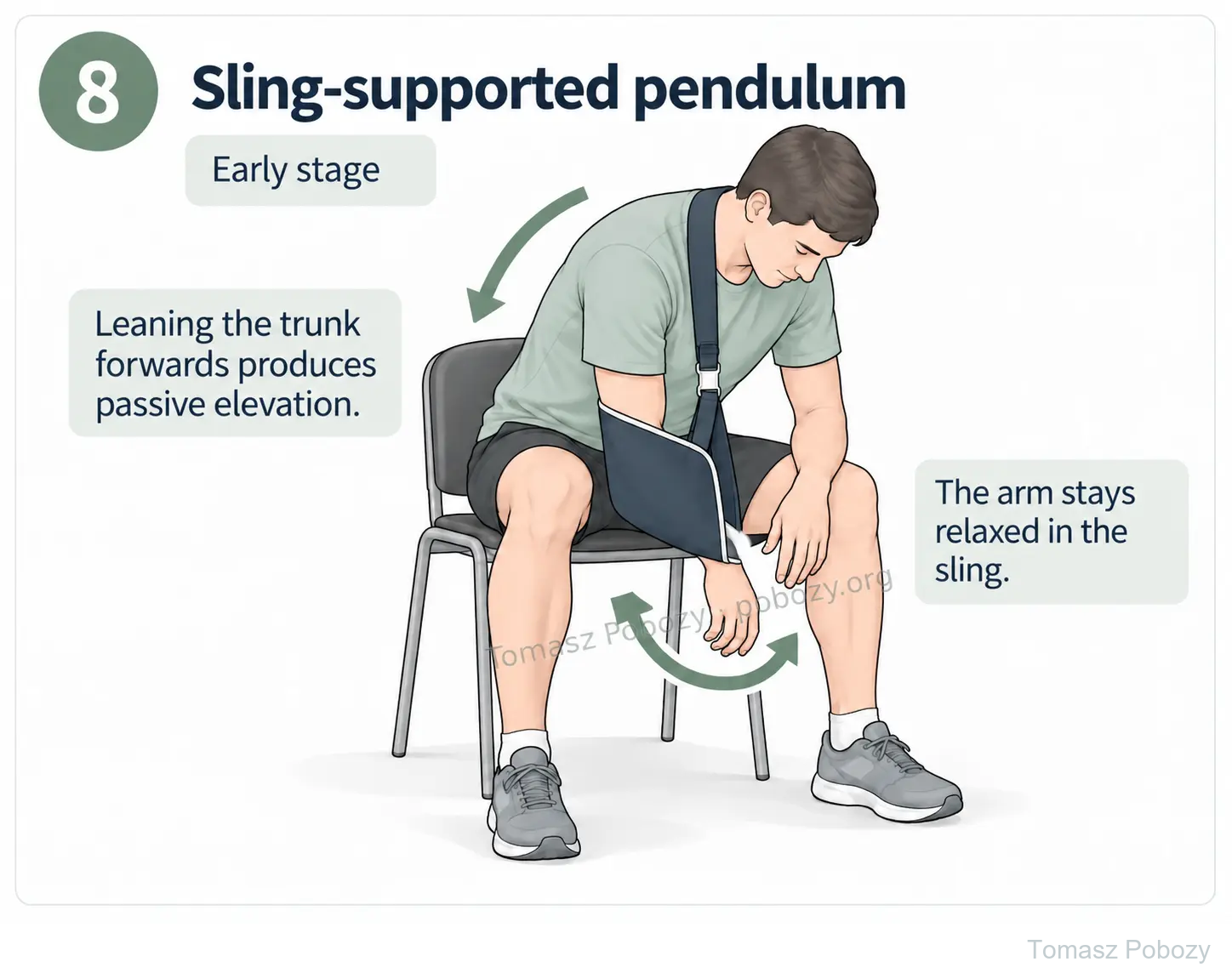
In some cases this may produce several tens of degrees of arm elevation relative to the trunk, sometimes approximately 60°. I treat this as an example of a possible range, not a mandatory target or a measure of isolated glenohumeral movement. This is my proposed modification, not a universal exercise variation assessed in the cited studies.
Passive movement must be permitted and appropriate support provided. If the arm is fixed to the trunk and leans with it, leaning alone does not increase shoulder flexion. A prescribed orthosis or stabilising strap must not be undone to gain more movement. The sling position and feasibility of the exercise must first be checked with the physiotherapist against the surgeon’s instructions.
The individual case, the individual patient, the individual surgeon.
Cooperation allows justified modifications and sometimes a shorter period of certain restrictions. When information supporting individual adjustments is unavailable, we follow the appropriate protocol and discharge instructions. At the same time, the physiotherapist continues to select exercise technique and dosage, assess progress and identify when the plan needs to change.
Use of this protocol
© 2026 Tomasz Poboży – orthopaedic surgeon. All rights reserved in the original synthesis and illustrations.