 Skip to content
FOR CLINICIANS AND PHYSIOTHERAPISTS
Subscapularis tendon repair — rehabilitation protocol
Protection, movement and a gradual return to daily activity. Select the stage appropriate to the actual procedure and current clearance.
Using and loading the limb
Early protection: no active or resisted internal rotation, lifting, pushing, pulling or body-weight support. Subscapularis resistance generally begins around week 12.
Full guidance ↓Brace or immobilisation
In this protective pathway, use a sling/brace for about 6 weeks, removing it for hygiene and prescribed exercise.
Full guidance ↓Permitted movement
Initially passive motion. ER progresses from neutral 0° toward about 30° by the end of week 6; large or tight repairs may require less range.
Full guidance ↓Permitted movement, loading and removal of a brace are separate decisions. Additional repairs may require a more protective programme.
Rehabilitation at a glance
Progression follows healing and functional criteria, not dates alone.
Rehabilitation boards
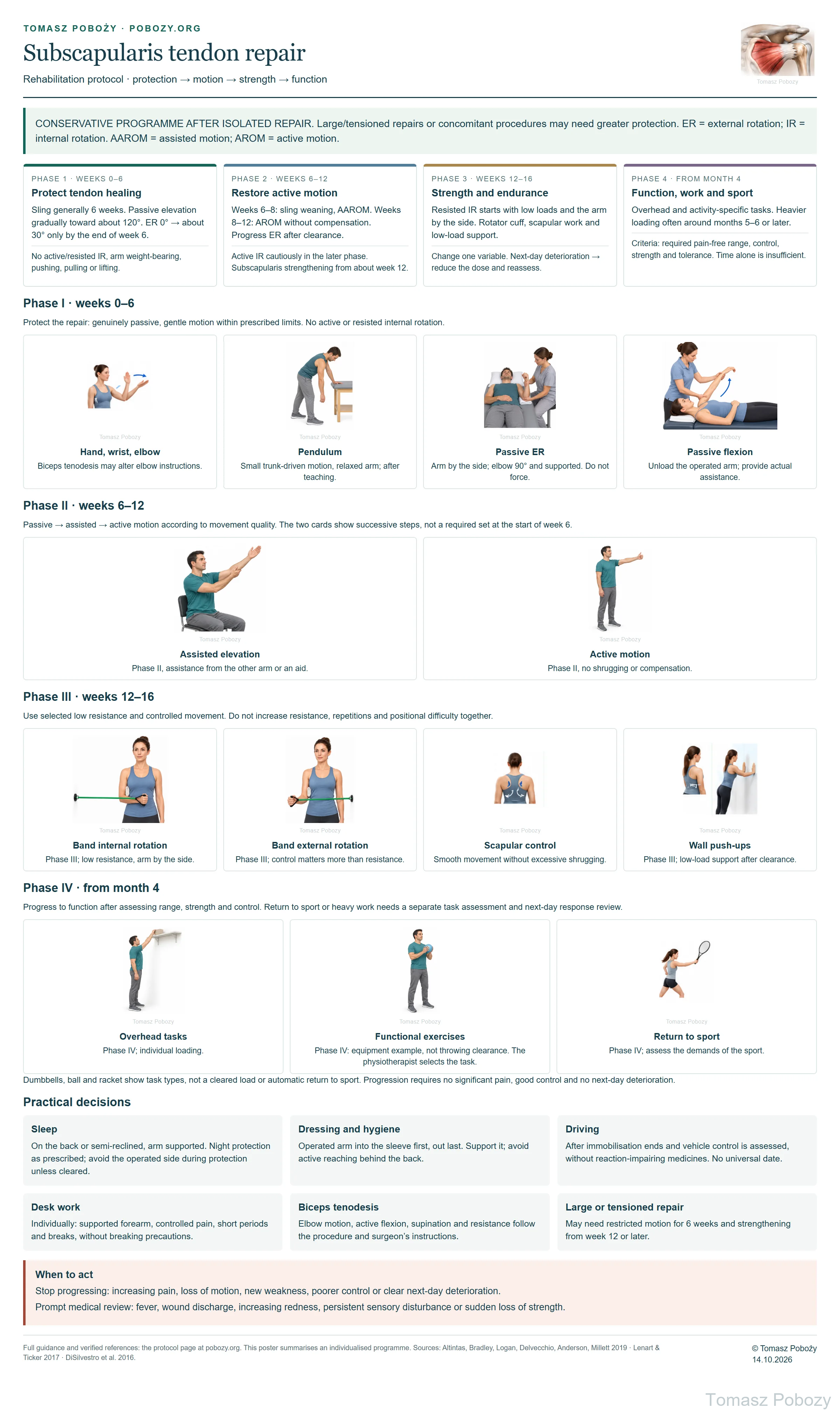
External rotation during the first 6 weeks: progress gradually from 0° to approximately 30° by the end of week 6. The detailed page text takes precedence over the graphic summary.
Exercises — a closer look
Select the phase allowed for your repair. Images do not authorise progression.
Phase I · 0–6
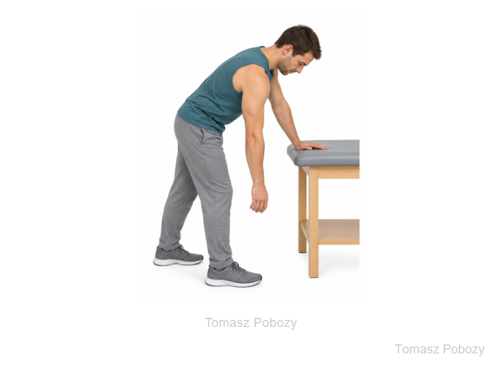
Relax the arm; trunk-driven movement, without a weight.
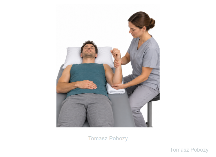
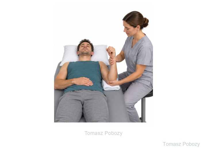
Elbow by the side, support and surgeon-prescribed limit.
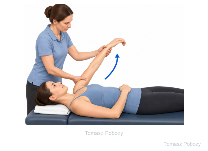
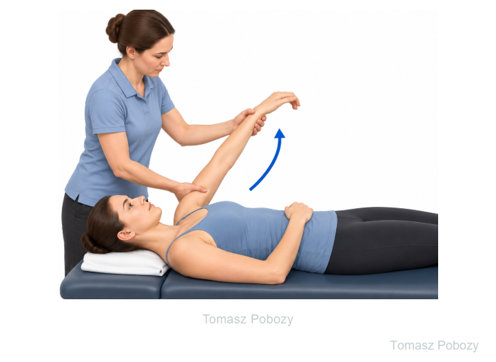
The therapist supports the arm; the patient does not actively lift it.
Phase II · 6–12
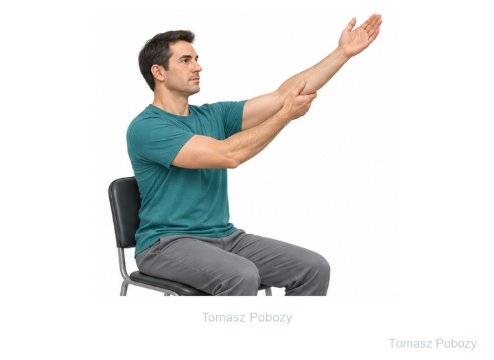
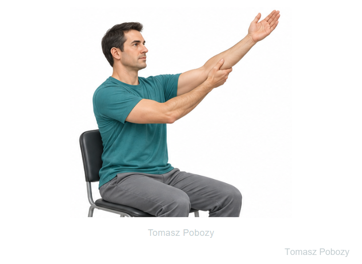
After clearance, with help from the other arm.
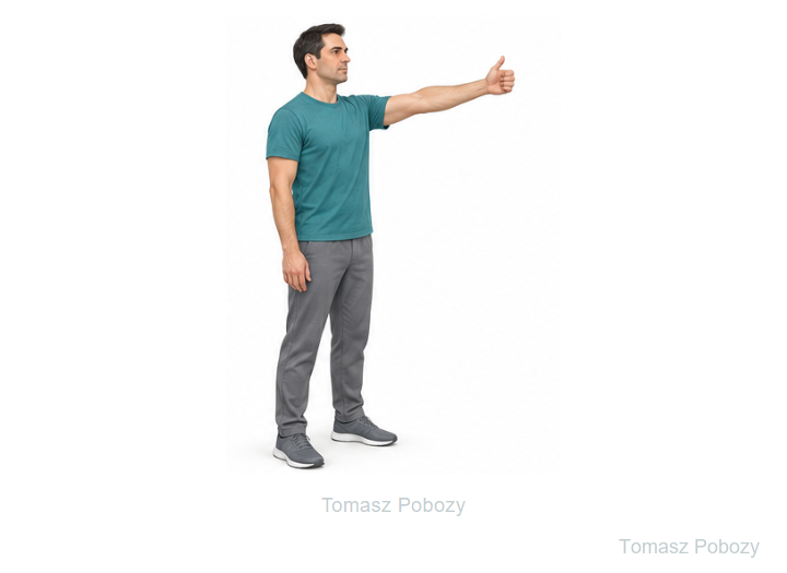
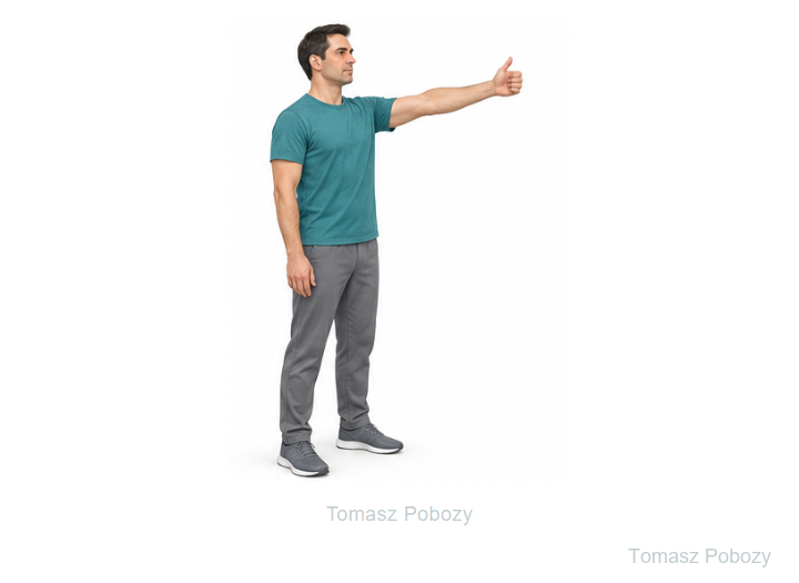
Next step after movement assessment, without a weight.
Phase III · 12–16
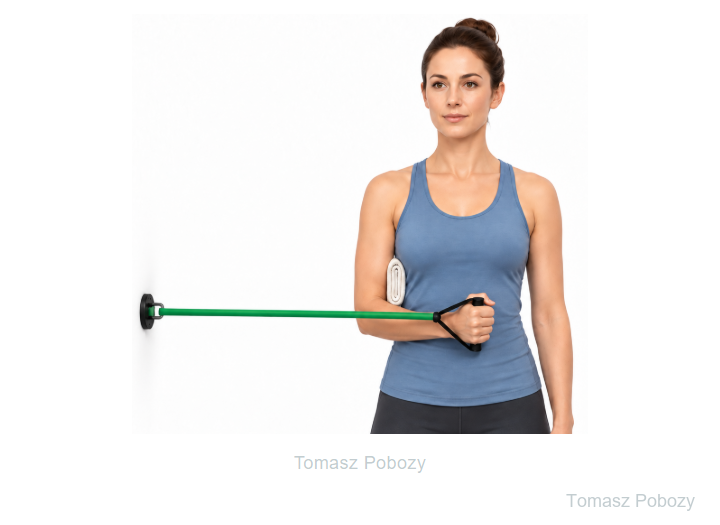
Low resistance at the side, only after clearance.
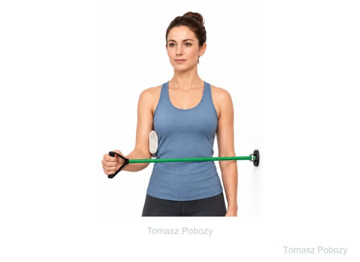
Control the motion; change one training variable at a time.
Phase IV · ≥4 mies. / months
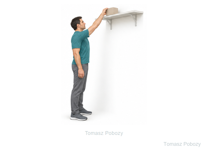
After motion, strength and control assessment; individualised load.
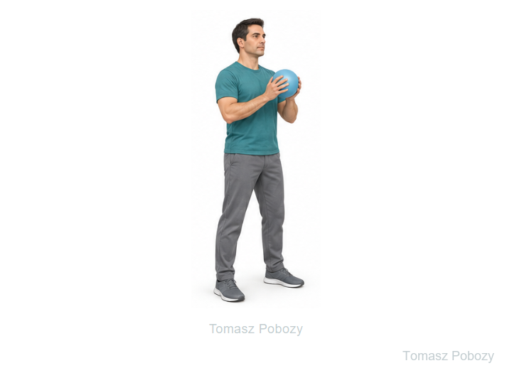
A task example, not clearance for throwing or a fixed load.
Scope and protection
Large repairs and concomitant procedures
A small upper-subscapularis repair may allow faster protected progression. A large/full-thickness tear, poor tendon quality or a repair under tension requires a more conservative approach. Concomitant biceps tenodesis or another repair introduces its own restrictions; follow the more protective pathway.
sling and protection
gradually 0° → 30° by the end of week 6
protect the subscapularis
generally from ~12 weeks
Stages and progression criteria
Phase I · 0–6 weeks — protection and passive motion
Goals: protect tendon-to-bone healing, control pain, maintain hand/wrist/elbow motion and preserve safe shoulder mobility.
Shoulder: passive motion only. For an uncomplicated repair, elevation is progressed gradually toward approximately 120°. External rotation starts at neutral (0°) and is increased progressively over the first 6 weeks, reaching approximately 30° only toward the end of week 6. Thirty degrees is the end-of-phase ceiling, not an early target.
Avoid: active or resisted internal rotation, forceful hand-behind-back stretching, weight bearing through the operated arm, pushing, pulling and lifting.
Phase II · 6–12 weeks — restoring active motion
Wean from the sling. Progress passive → active-assisted → active motion according to movement quality. External rotation is increased gradually beyond the previous limit if the orthopaedic surgeon has not specified otherwise; aggressive stretching is avoided.
Active internal rotation is introduced cautiously in the later part of this phase. Substantial strengthening of the repaired subscapularis is generally deferred until about 12 weeks.
Phase III · 12–16 weeks — strength and endurance
Begin resisted internal rotation with low resistance and the arm by the side. Progress resistance, repetitions and arm position gradually. Strengthen the remaining rotator cuff and scapular stabilisers and introduce closed-chain loading from low levels such as wall push-ups.
Change one loading variable at a time. Increasing pain, loss of movement quality or a clearly worse shoulder the following day means returning to the previous dose.
Phase IV · from 4 months — function, work and sport
Progress to greater resistance, overhead work and activity-specific tasks. Full return to heavier work or sport often occurs around 5–6 months or later and should be based on functional range, good scapulohumeral control, acceptable strength and load tolerance rather than time alone.
Daily activities
Support the arm during sleep and hygiene; sling use follows the postoperative instructions. Dress the operated arm first and undress it last. Driving requires sling discontinuation, safe vehicle control and no impairing medication. Desk work uses forearm support and breaks. Heavy work and sport require separate functional assessment; the start of month four is not clearance for full activity.
Warning signs
Call 112 for sudden breathlessness, chest pain or fainting.
Warning signs
Stop progression and contact the surgical team for uncontrolled or increasing pain, a fever, excessive wound drainage, persistent numbness or tingling, or a sudden loss of previously recovered function. Do not test the repair with additional resistance. Mass General Brigham — Subscapularis Repair [historical link — PDF currently unavailable at this address]
For a life-threatening emergency, call your local emergency number immediately — 112 in the European Union. On the boards, 112 refers to the EU.
Additional scope notes
SHOULDER AND UPPER ARM · FOR DOCTORS AND PHYSIOTHERAPISTS
Protection of the repair from excessive external rotation and premature loading of internal rotation, followed by a staged return of motion, strength and function.
Prepared by Tomasz Poboży, MD, PhD — orthopaedic surgeon
The poster is a summary. Detailed restrictions and progression criteria are described below. Select the poster to open the full image.
References
References
- Altintas B, Bradley H, Logan C, Delvecchio B, Anderson N, Millett PJ. Rehabilitation Following Subscapularis Tendon Repair. Int J Sports Phys Ther. 2019;14(2):318–332. DOI: 10.26603/ijspt20190318
- Barlow JD, Everhart JS. Arthroscopic Subscapularis Repair Through a Single Anterior Portal. Arthrosc Tech. 2017;6(5):e1593–e1598. DOI: 10.1016/j.eats.2017.06.014
- You JS, Monroe EJ, Friedman JM, Feeley BT, Lansdown DA, Zhang AL, Ma CB. Arthroscopic Single-Portal Subscapularis Tendon Repair. Arthrosc Tech. 2020;9(10):e1447–e1452. DOI: 10.1016/j.eats.2020.06.005
- Lenart BA, Ticker JB. Subscapularis tendon tears: Management and arthroscopic repair. EFORT Open Rev. 2017;2(12):484–495. DOI: 10.1302/2058-5241.2.170015
- Mass General Brigham. Rehabilitation Protocol for Subscapularis Repair. Wersja oznaczona Revised 10/2022 w zaindeksowanej treści. Historical institutional document. The former PDF URL currently redirects to a general hospital page; it is not a verified current basis for the timetable.
The Altintas paper is a clinical commentary (level 5). Technical reports describe the operation; they do not validate this entire rehabilitation schedule. This page uses the author’s protective variant.
Use of this protocol
Educational material for doctors and physiotherapists. Application requires review of the operative report, the individual patient’s condition and the surgeon’s postoperative instructions.
© 2026 Tomasz Poboży – orthopaedic surgeon · pobozy.org