 Skip to content
FOR CLINICIANS AND PHYSIOTHERAPISTS
Bankart repair with anchors — rehabilitation protocol
From protecting the repaired capsulolabral complex to a safe return to work and sport. Two boards present the rehabilitation stages, movement limits and examples of progression. Expandable details, commentary and references follow below.
Using and loading the limb
During protection, no lifting, sudden traction or body-weight support. Resistance through a larger range generally follows 6 weeks and clearance.
Full guidance ↓Brace or immobilisation
Use the prescribed sling or brace, usually about 3 weeks in this pathway. Do not shorten a longer prescribed period.
Full guidance ↓Permitted movement
Initially, external rotation only to neutral. Avoid combined abduction and ER and reaching behind the back; further range follows the repair-specific plan.
Full guidance ↓Permitted movement, loading and removal of a brace are separate decisions. Additional repairs may require a more protective programme.
Rehabilitation at a glance
Progression follows healing and functional criteria, not dates alone.
Rehabilitation boards
TOMASZ POBOŻY · REHABILITATION PROTOCOL
Bankart repair · protection and weeks 0–6
Primary arthroscopic soft-tissue repair with anchors. Bone procedures, revision repairs and additional procedures require separate consideration.
BEFORE STARTING
Know the repair
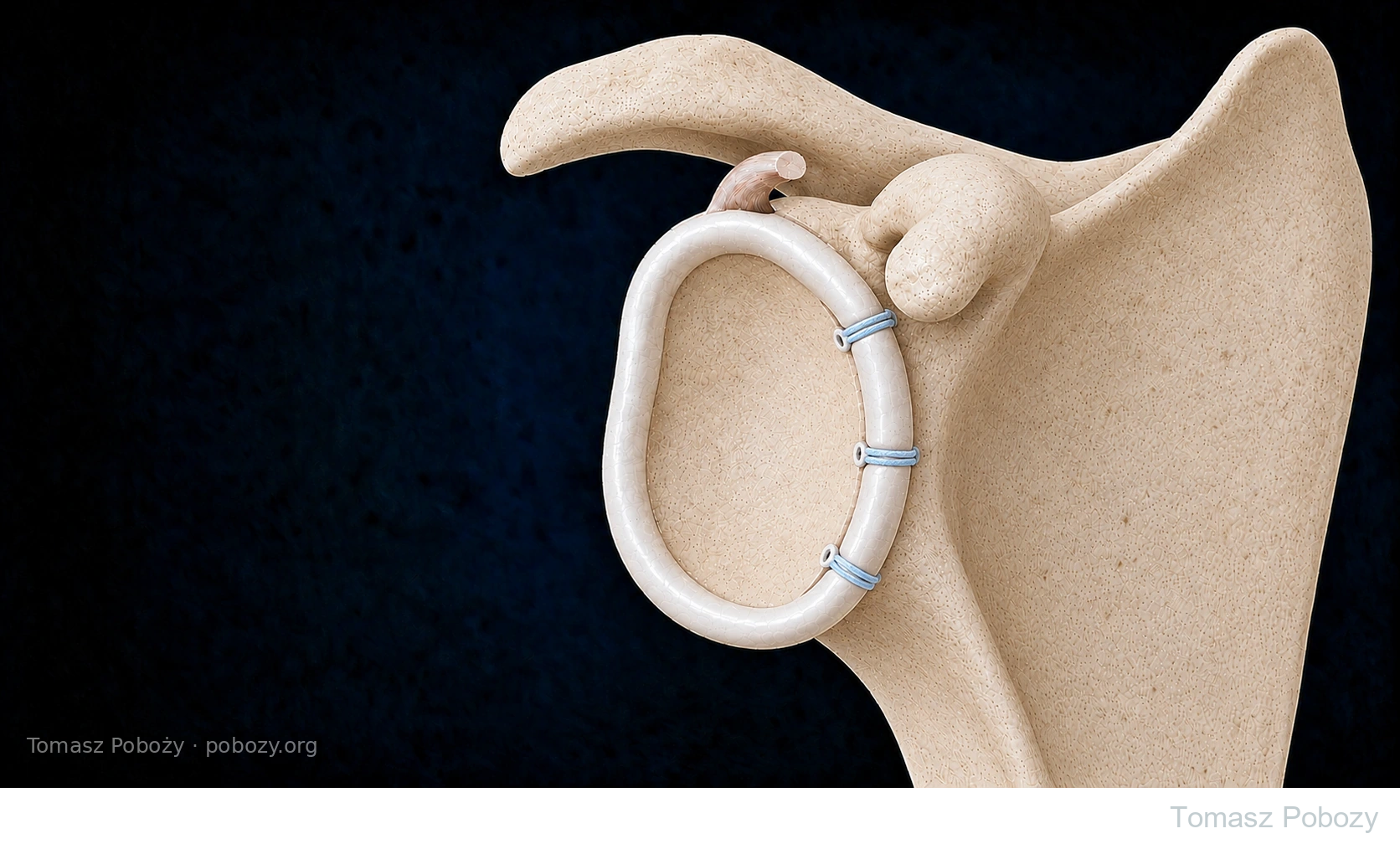
Read the operative report and discharge instructions: extent of repair, capsular shift, tissue quality, concomitant procedures, immobilisation and permitted movement.
Regular and as-needed pain medication follow their prescribed schedules. Do not suppress pain to force range.
01 · WEEKS 0–3
Protect the repair
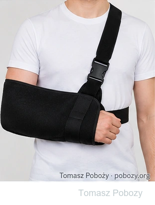
Use the prescribed sling or orthosis. Delphi consensus generally describes protection for about three weeks; do not shorten a longer surgeon-prescribed period.
Protected movement limits in the BESS synthesis: elevation up to 90° anterior to the scapular plane; external rotation to neutral (0°).
These are limits during immobilisation, not targets to force in the first session. Exercise the hand, wrist and elbow as instructed.
02 · ABOUT WEEKS 3–6
Movement & controlled activation
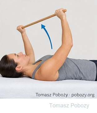
After clearance, gradually reduce sling use and increase movement. Supported and assisted movement leads towards smooth active control.
Introduce gentle isometrics and scapular control as selected by the physiotherapist. Isometrics still load tissues.
No resistance exercises through a larger range yet. External rotation is progressed cautiously and depends on arm position.
PROTECTION THROUGHOUT
Avoid early stress
Avoid combined abduction and external rotation, reaching behind the back, lifting, sudden pulls and body-weight support through the operated hand.
Do not force end range or movements that produce a sense of slipping. Permitted movement is not clearance for loading.
Summary of the approved protocol. Read the detailed sections below. The individual repair and postoperative instructions take priority. © 2026 Tomasz Poboży — orthopaedic surgeon.
TOMASZ POBOŻY · REHABILITATION PROTOCOL
Bankart repair · strength and return to activity
Progress by healing, function and task readiness. The start of a new phase does not itself clear contact sport or unrestricted loading.
03 · ABOUT WEEKS 6–12
Strength, endurance & control
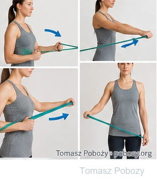
After assessment, start movement against light resistance, then increase range and difficulty. Train rotators, scapular muscles and coordination with the trunk.
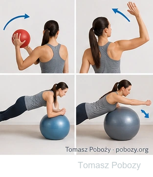
Once arm support is permitted, begin small controlled weight shifts in an easy position, such as at a wall. Avoid early deep push-ups, dips, fly exercises and aggressive anterior stretching.
04 · AFTER WEEK 12
Prepare for work & sport
Build endurance and control with increasingly task-specific exercises. Add range, resistance, speed and unpredictability progressively.
Throwing, overhead activity and rapid deceleration require separate clearance. High-load return may be at 4–6 months or later; this is not a guaranteed deadline.
05 · WHEN CRITERIA ARE MET
Return to full activity
Combine healing, the orthopaedic surgeon’s assessment, functional testing and patient confidence. Assess required range, rotator strength and endurance, support and dynamic control.
A symmetry percentage alone is insufficient. Late testing positions must not be used to challenge a fresh repair.
Selected tasks → partial training → full training → competition or full duties. Consider contact and fall risk separately.
STOP & REASSESS
Warning signs
New slipping, sudden pain after injury, lost function, fever, wound discharge or persistent altered sensation needs assessment.
Suspected redislocation requires urgent medical attention. Do not progress just because another week has passed.
Summary of the approved protocol. Read the detailed sections below. The individual repair and postoperative instructions take priority. © 2026 Tomasz Poboży — orthopaedic surgeon.
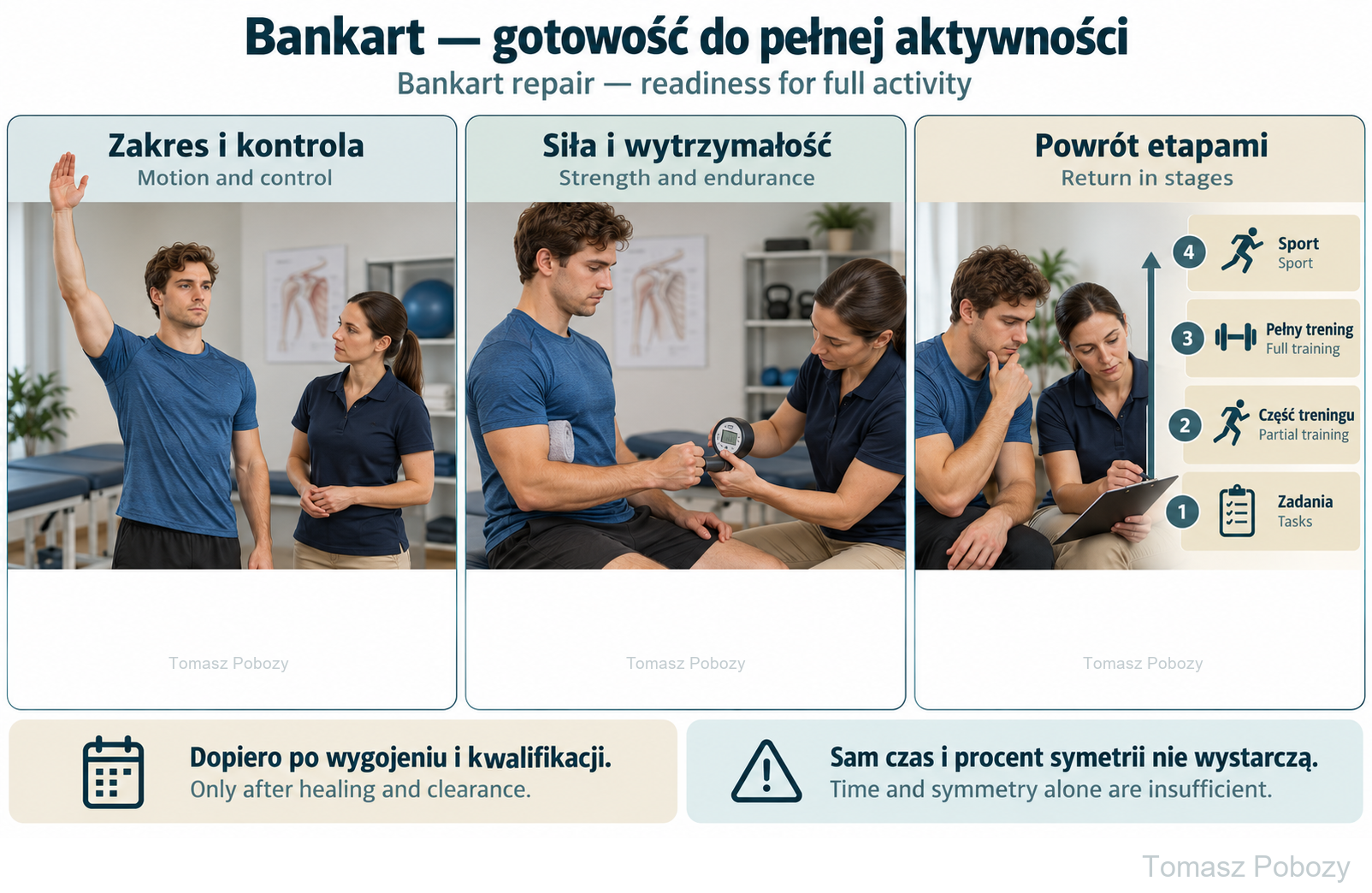
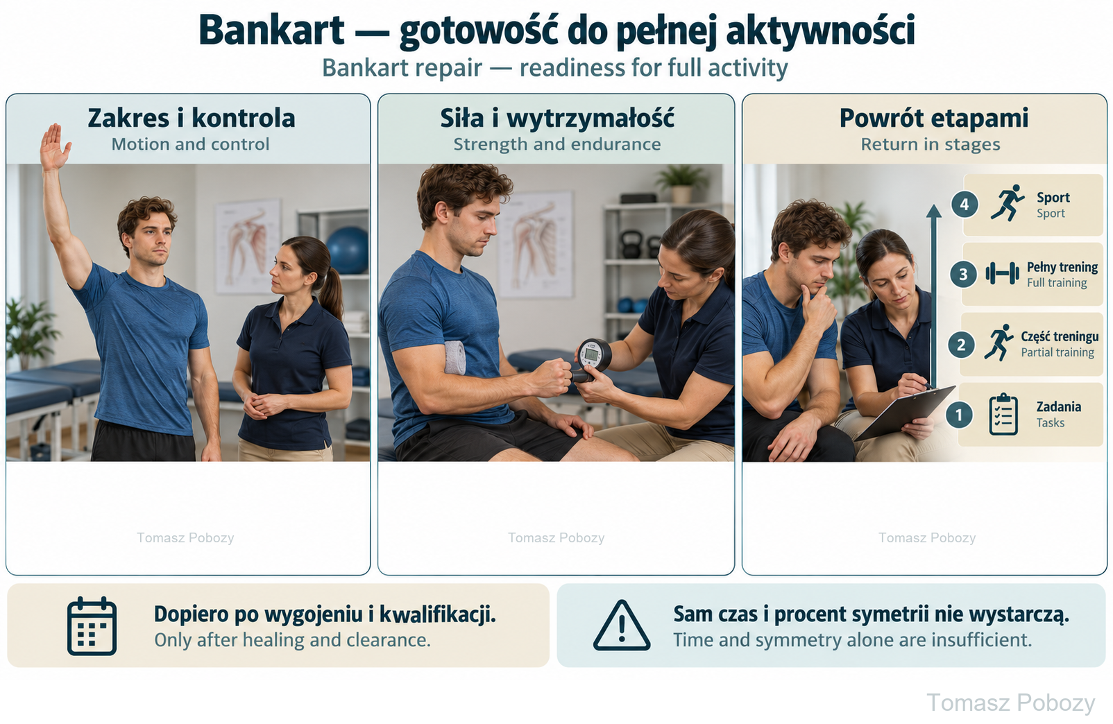
Assess motion, control, strength and endurance after healing and clearance. The strength-measurement illustration represents clinician assessment, not a home test or an exercise for a fresh repair. Progress from selected tasks to partial training, full training and sport. Time and symmetry alone are insufficient.
Exercises — a closer look
Use the complete boards above together with the instructions for the chosen stage. An exercise shown for a later stage is not permitted early.
Assess motion, control, strength and endurance after healing and clearance. The strength-measurement illustration represents clinician assessment, not a home test or an exercise for a fresh repair. Progress from selected tasks to partial training, full training and sport. Time and symmetry alone are insufficient.
Open the relevant complete board and zoom in. The viewer retains all stages and captions.
Exercise instructions and progression are contained in the stage descriptions linked above.
Scope and protection
Scope and protection of the repair
When does this protocol apply?
After primary arthroscopic anchor repair of the anteroinferior labrum and adjacent capsule in an adult with traumatic anterior instability. The extent of capsular shift and tension must be reflected in the postoperative instructions.1, 2
Outside the scope: Latarjet, bone grafts and bone-block procedures, fixation of a bony Bankart lesion, revision and open repairs. The pathway does not automatically cover additional remplissage, posterior or extensive circumferential labral repair, SLAP repair or rotator cuff repair. Remplissage also requires separate consideration, even though it is not a bone procedure.6
Start with the discharge summary and operative report
Information is needed on the extent of repair, concomitant procedures, the type and duration of immobilisation, and permitted movement and loading. “Bankart repair with anchors” alone is not enough to set the pace of rehabilitation. Unclear instructions should be clarified with the surgeon.
Pain control from the first days
Analgesics prescribed for regular use are taken according to the written schedule; medication prescribed as needed remains as needed. The aim is to allow rest and permitted exercises, not to suppress pain in order to force movement. Cooling, if prescribed, is used with skin protection and after sensation has returned following a nerve block.
Exercises and home programme
Exercise examples and home programme
The descriptions below explain technique; they are not a set to perform from day one. The physiotherapist selects exercises appropriate to the phase and teaches the patient to reproduce them correctly.
Supported table slide
The arm rests on a towel on a table. A small forward trunk lean and assistance from the other hand help movement without placing body weight through the operated limb. During initial protection, movement stays within the permitted zone.2
Stick-assisted movement
The unaffected hand guides the operated arm through a light stick. For rotation exercises, the elbow is supported and remains close to the trunk. The stick is not used to force through resistance. Rotation beyond neutral requires prior permission to expand the range.2
Daily home work, with strengthening dosage set separately
Physiotherapy sessions are supplemented by a daily, individually prescribed home programme. It includes permitted mobility and arm control; resistance exercise frequency and recovery intervals are set separately. The same number of visits, sets and repetitions is not imposed on everyone. The patient should receive clear instructions on what to do, within what range, how often and when to stop.
Regaining confidence in the shoulder
After previous dislocations, recovery of range and strength does not always remove apprehension about movement. The physiotherapist gradually introduces harder tasks that have already been cleared. REGUIDE emphasises education, graded exposure and whole-kinetic-chain exercises. This does not mean provoking early instability or dismissing actual shoulder slipping as anxiety alone.4
When should progression stop?
A new sense of the shoulder slipping, sudden pain after a jerk or injury, loss of previously gained function, fever, wound discharge or persistent altered sensation requires assessment. Exercise difficulty must not be increased merely because another week has passed. Suspected redislocation requires urgent medical attention.
Stages and progression criteria
Phases I–II: surgery to week 6
Stages of rehabilitation
The stages cover protection during the first three weeks, mobilisation and early muscle activation during weeks 3–6, strengthening during weeks 6–12, and later return to sporting tasks and high loads. Time since surgery remains important, but does not replace functional assessment.1
PHASE I · WEEKS 0–3
Protecting the repair and moving within the permitted range
Immobilisation: the sling or orthosis prescribed at discharge. The Delphi consensus generally provides for sling protection for approximately three weeks; a longer period prescribed by the surgeon must not be shortened independently.3
up to 90°
of arm elevation anterior to the scapular plane
up to 0°
of external rotation — to neutral
These are limits of protected movement during immobilisation described in the BESS synthesis, not values that must be reached in the first session. With the arm at the side, neutral rotation means the forearm points forwards with the elbow bent to 90°. In this phase, the forearm is not moved farther outwards.2
Within the protected range, passive, assisted and permitted active movement may be used after instruction. The hand, wrist and elbow are also exercised. The timing and method of movement follow the instructions for the repair.2, 3
Avoid: combining abduction with external rotation, reaching behind the back, lifting, jerking and supporting body weight through the operated hand. Permitted movement does not mean clearance for loading.
Before progressing: the patient understands the restrictions, performs the taught movements without a clear symptom flare, and the wound and clinical course raise no concerns.
PHASE II · APPROXIMATELY WEEKS 3–6
Gradually increasing movement and controlled activation
Once cleared by the surgeon, sling use is gradually reduced and range of motion increased. Assistance from the other hand and support of the limb help the transition to smooth independent movement. Scapular control and gentle muscle contractions in protected positions are developed.1
An isometric exercise involves contraction without visible movement, but still loads tissues. The physiotherapist selects the direction and intensity. Resistance exercises through a larger range of motion are not yet introduced. The consensus places this type of strengthening after six weeks, rather than at an arbitrarily earlier time.3
External rotation is advanced particularly carefully. Clearance for greater rotation with the arm at the side does not permit the same movement with the arm abducted. End ranges and movements producing a sense of the humeral head slipping must not be forced.
End-of-phase goal: greater independence in permitted activities and controlled movement within the range set for the repair, without pursuing full mobility at any cost.
Phases III–V: week 6 to full activity
PHASE III · APPROXIMATELY WEEKS 6–12
Rebuilding strength, endurance and control
After clinical assessment, movement against light resistance begins, with range and difficulty then increased. The programme includes shoulder rotators, scapular control muscles and coordination of the arm with the trunk. Early examples include rotation with a light band and the elbow close to the body, and scapular exercises without taking the arm far backwards.1, 3
Once weight-bearing through the arm is cleared, small controlled weight shifts are introduced, initially in an easy position such as against a wall. Deep push-ups, fly exercises, dips and sudden overhead movements are not starting exercises. Aggressive anterior capsular stretching is avoided.
End-of-phase goal: a functional, well-controlled range and tolerance of the selected resistance. Strength gains should not come at the expense of movement quality or increasing feelings of instability.
PHASE IV · AFTER WEEK 12
Preparing for work and sport
Exercises increasingly resemble the patient’s actual tasks. Training develops endurance, control during changes of direction and coordination of the shoulder, trunk and lower limbs. Greater range, resistance, speed and unpredictability are introduced gradually rather than all at once.1, 4
Starting this phase does not mean clearance for unrestricted contact sport. Throwing, overhead movement and loads requiring rapid deceleration of the arm need separate clearance. Return to high loads may occur at 4–6 months or later, but this is not a guaranteed date for everyone.
Goal: performing progressively harder work or sport tasks without poorer tolerance of subsequent sessions.
PHASE V · WHEN CRITERIA ARE MET
Return to full activity
The decision combines the course of healing, the surgeon’s assessment, functional examination and the patient’s readiness. Assessment covers the range needed for the activity, rotator strength and endurance, control during support and dynamic movement, and confidence in performing tasks. Testing in abduction with external rotation belongs to late assessment, not to testing a fresh repair.3, 4
Strength should be measured under reproducible conditions, for example with a dynamometer, accounting for arm dominance and the other shoulder’s condition. A single symmetry percentage is not independent clearance to return. A 2025 study found that even healthy volunteers failed some proposed tests and thresholds; results must be interpreted in clinical context.5
Return sequence: selected training tasks → partial training → full training → competition or full work duties. Contact level and fall risk are considered separately.
Daily activities and follow-up
Daily activities follow the permitted shoulder and elbow motion, lifting and arm support for your procedure. Sling removal is not clearance for lifting or driving. The detailed guidance below explains the criteria and exceptions.
From protecting the repaired capsulolabral complex to a safe return to work and sport. Two boards present the rehabilitation stages, movement limits and examples of progression. Expandable details, commentary and references follow below.
Warning signs
Warning signs
For a life-threatening emergency, call your local emergency number immediately — 112 in the European Union. On the boards, 112 refers to the EU.
Additional scope notes
The boards summarise the stages of rehabilitation. Open the sections below for the full description.
References
Scientific basis and references
Scientific basis and references
The phase framework comes from the 2026 BESS recommendations; early movement limits come from the synthesis published in the British Journal of Sports Medicine. The Delphi consensus and REGUIDE supplement the description of loading, function and exercise examples. This is a synthesis for a defined group of procedures, not a complete translation of one protocol or a separately validated programme. Strong comparative evidence is still lacking for many rehabilitation details.1, 2, 3
- Wong C, Jaggi A, Kearney R, Gwilym S. British Elbow and Shoulder Society practice guidelines: Rehabilitation following traumatic anterior shoulder dislocation (post-operative and non-operative care). Shoulder & Elbow. 2026;18(3):604–608. doi:10.1177/17585732261439731. The postoperative care section was used.
- Wong C et al. Critical evidence synthesis on rehabilitation following arthroscopic shoulder stabilisation surgery for traumatic anterior instability: consensus recommendations for clinical practice and research – commissioned by the British Elbow & Shoulder Society. Br J Sports Med. 2026;60(2):98–107. doi:10.1136/bjsports-2025-109674. Protected range described on the basis of the abstract.
- Willmore E et al. Rehabilitation guidelines following arthroscopic shoulder stabilisation surgery for traumatic instability – a Delphi consensus. Physiotherapy. 2024;124:154–163. doi:10.1016/j.physio.2024.05.001. Expert consensus; agreed recommendations should be distinguished from proposals that did not achieve consensus.
- Marianne Larsen van Gastel et al.; International Shoulder Instability Group. Developing a Rehabilitation Guideline (REGUIDE) for Patients Undergoing an Arthroscopic Bankart Repair After Traumatic Anterior Shoulder Dislocation, Focusing on Managing Apprehension: An International Delphi-Based Consensus Study. J Orthop Sports Phys Ther. 2024;54(5):289–301. doi:10.2519/jospt.2024.12106. Main principles discussed on the basis of the abstract.
- Hargreaves M et al. A High Percentage of Healthy Volunteers Fail to Pass Criteria-based Return to Sport Testing for Arthroscopic Bankart Repair. Arthroscopy. 2025;41(8):2785–2791. doi:10.1016/j.arthro.2025.01.047. Discussion based on the abstract.
- Villarreal-Espinosa JB et al. Rehabilitation Protocol Variability Following Arthroscopic Bankart Repair and Remplissage for Management of Anterior Shoulder Instability: A Systematic Review. Int J Sports Phys Ther. 2024;19(10):1172–1187. doi:10.26603/001c.123481. Context for separate management after remplissage, not a programme for isolated Bankart repair.
Clinical commentary and terms of use
Commentary
Commentary
The same procedure name does not mean the same repair
When I hear “Bankart repair”, my first concern is what was actually reconstructed. Securely reattaching a preserved labrum to the glenoid rim differs from reconstructing a structure to take over its function from a weakened or altered ligamentolabral complex. The extent of injury, tissue quality, repair tension and bone condition also matter.
Anchor fixation alone does not establish that both situations permit identical rehabilitation. A good-quality repair may justify earlier clearance for selected activities, while poorer quality may call for longer protection. I would not turn this observation into a new table with automatically assigned weeks. Individualisation concerns the individual case, patient and surgeon.
Cooperation between surgeon and physiotherapist
The physiotherapist helps shape the rehabilitation plan, selecting exercises and dosage and assessing movement quality, loading tolerance and functional recovery. The surgeon contributes knowledge of tissue condition and the repair achieved. Information should flow in both directions — not only about problems, but also about progress and proposed programme changes.
We do not test the durability of a fresh repair with a strength challenge or by provoking apprehension about dislocation. The operative documentation or a discussion with the surgeon is needed. When there is no basis for agreeing individual adjustments, we follow the appropriate protocol and discharge instructions. This does not remove the physiotherapist’s independence in selecting how to deliver therapy within the agreed limits.
A mobility target versus a protective limit
The desired range at the end of a phase is not a target to achieve at any cost. Protecting repaired tissues takes priority. However, this must be distinguished from a protective limit that applies during a particular period. The initial limits on elevation and external rotation are boundaries that must not be exceeded without a change in instructions, even if the patient could perform a larger movement.
External rotation is not assessed without reference to arm position. The record should state whether it was tested with the arm at the side, in the scapular plane or at a specified degree of abduction. Only measurements taken in comparable positions allow meaningful assessment of progress.
Use of this protocol
© 2026 Tomasz Poboży – orthopaedic surgeon. All rights reserved in the original synthesis and illustrations.