Autor: Tomasz Poboży

  • IVF szybciej niż inseminacja, lecz po 20 miesiącach wynik był podobny

    W wieloośrodkowym, randomizowanym badaniu FIIX uczestniczyło 737 par z niepłodnością niewyjaśnioną. Porównano do czterech cykli inseminacji domacicznej ze stymulacją jajników z jednym pełnym cyklem zapłodnienia pozaustrojowego (IVF). W pierwszym etapie, obejmującym siedem miesięcy, urodzenie dziecka odnotowano u 31% par w grupie inseminacji i 55% w grupie IVF. Gdy parom bez powodzenia umożliwiono dalsze leczenie i oceniono cały okres do 20 miesięcy, skumulowane wyniki były już bardzo podobne: 70% i 71%. To duże badanie kliniczne u ludzi sugeruje, że IVF może szybciej prowadzić do urodzenia dziecka, ale nie dowodzi, że każda para z niepłodnością niewyjaśnioną powinna rozpoczynać właśnie od tej metody; decyzja wymaga indywidualnego uwzględnienia wieku, obciążeń leczenia, kosztów i preferencji pary.

    Źródło: Prentice LR, Sadler LC, Anderson KL i wsp. New England Journal of Medicine, publikacja online 27.10.2026. DOI: 10.1056/NEJMoa2602728. Sprawdzono 30.10.2026; odczytano abstrakt indeksowany w PubMed i metadane NEJM (PMID 42950222). Pełny tekst nie był dostępny w użytym dostępie. W chwili kontroli nie znaleziono korekty ani wycofania pracy.

    ← Wróć do „Ze świata medycyny”

    Opracowanie: Tomasz Poboży - ortopeda.

  • Rotator Cuff Tears — When Is Surgery Needed?

    Rotator Cuff Tears — When Is Surgery Needed?

    PATIENT INFORMATION · SHOULDER

    We very often consider surgery for full-thickness tears, but it is not always necessary.

    The cause and size of the tear, tissue quality and shoulder function all matter. A recent, large traumatic tear with sudden weakness needs prompt assessment and may favour early repair.

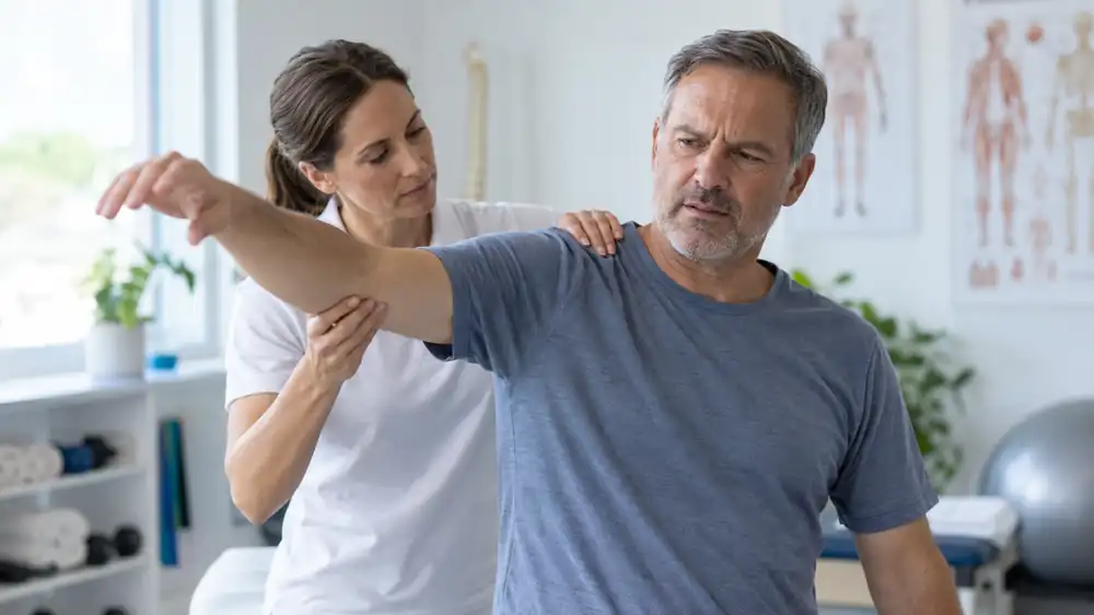
    Shoulder weakness during clinical assessment of a rotator cuff tear. Medical content prepared by: Tomasz Poboży - orthopaedic surgeon.

    PODCAST · DOCTOR POBOŻY ON HEALTH

    Rotator Cuff Tears — When Is Surgery Needed?

    An English episode about early repair after injury, individual rehabilitation, imaging and the role of injections.

    Listen on Spotify ↗

    What does a rotator cuff tear mean?

    The rotator cuff consists of the tendons of four muscles surrounding the head of the upper arm bone. They stabilise the shoulder and help lift and rotate the arm. Tears most often affect the supraspinatus tendon, sometimes extending into part of the infraspinatus tendon.

    A tear can involve part or all of the tendon’s thickness. It may occur suddenly after a fall or a forceful pull, but often develops gradually with degenerative tendon changes. Pain when raising the arm, weakness and difficulty sleeping on the affected side are common symptoms.

    An abnormal scan does not establish the source of every symptom. Some tears cause little pain; symptoms may also arise from the subacromial bursa, the long head of the biceps tendon, stiffness or arthritis. Findings need to be matched to the examination.

    When is earlier surgery preferable?

    A recent, large traumatic tear, especially with sudden weakness or inability to lift the arm, usually favours early surgical repair. If the tendons are repairable and the patient’s health permits surgery, there is no need to complete months of rehabilitation first.

    Prompt orthopaedic assessment is important. Tendon retraction, muscle wasting and fatty changes can make later repair more difficult. The timing is planned individually after assessment; this does not mean that every tear needs emergency surgery on the day of injury.

    Other reasons to consider surgery include persistent pain, weakness and functional loss despite appropriate nonsurgical treatment, a repairable tear at risk of progression, and substantial work or sports demands. No single age or tear-size threshold settles the decision for every patient. [1, 2]

    When can treatment start without surgery?

    For many gradually developing tears, particularly smaller ones without major loss of function, individual rehabilitation is a reasonable starting point. This also applies to some symptomatic full-thickness tears. Both physiotherapy and surgery can improve outcomes in small and medium full-thickness tears. [1]

    Treatment involves adapting activities that worsen pain, restoring movement, improving rotator cuff and shoulder blade muscle strength and control, and gradually increasing loading. It is an active plan, not simply avoiding use of the arm. Pain medicines are selected individually, taking contraindications into account.

    Pain relief does not necessarily mean the tendon has healed. Tear size, muscle wasting and fatty changes may progress in some patients, so symptom improvement and tendon condition both need follow-up. [1, 3, 4]

    What do longer-term studies show?

    In the prospective MOON cohort of 452 patients with symptomatic, atraumatic full-thickness tears, more than 70% were managed successfully without surgery. Patient-reported outcomes improved in the first months and did not decline over ten years among those continuing nonsurgical care. These findings do not mean that every tendon healed or that surgery is unnecessary for every tear.

    Long-term comparative research also supports the importance of repair in selected patients and monitoring of tear progression. Conservative management means reviewing function, symptoms and tissue condition rather than leaving the problem unassessed for years. [3, 4]

    Ultrasound, MRI or X-ray?

    Assessment starts with the history and examination. Combining examination findings is more useful than relying on one provocative test. Imaging should answer a clinical question rather than replace the examination. [1]

    What can ultrasound show?

    Ultrasound assesses tendon continuity, the location and extent of many tears, the biceps tendon and bursa. Selected structures can be observed during movement. It is a useful examination when performed and interpreted by an experienced clinician.

    When is MRI useful, and what do X-rays add?

    MRI helps assess tendon retraction, muscle wasting and fatty changes, and deeper shoulder structures. It is particularly useful for surgical planning or when examination and ultrasound leave important questions unanswered. X-rays add information about bones, joint alignment and arthritis.

    Injections: different treatments, different aims

    Corticosteroid: an injection into the subacromial bursa may reduce inflammation and pain, but does not repair a torn tendon. Repeated injections require caution, particularly when tendon repair is planned. They should not be used to indefinitely postpone indicated surgery. [1]

    Platelet-rich plasma (PRP): in selected partial tears, including splits within the tendon, treatment may be considered with the aim of supporting repair processes. Results are mixed; routine PRP use for partial tears is not supported by the AAOS guideline. Improvement in symptoms should not be equated with proven structural healing. [1]

    Injectable platelet-rich fibrin (i-PRF): this is also being investigated, but evidence in these injuries remains limited. Published case reports involving liquid fibrin do not establish its superiority to PRP. Neither blood-derived treatment replaces surgery when surgery is needed. [5]

    How is the tendon repaired?

    Repair is most often arthroscopic, through small incisions. The tendon is secured to its attachment on the upper arm bone using anchors and sutures. The aim is to restore the attachment without excessive tension. The configuration, number of anchors and single- or double-row technique depend on tear size and shape, tendon quality and how far the tissue can be mobilised.

    Healing matters: healed repairs are associated with better functional outcomes than unhealed repairs and, in comparative studies, physiotherapy alone. Tissue quality, repair tension and biological healing potential therefore influence planning. [1, 2, 4]

    Rehabilitation after repair

    The repaired tendon needs protection and gradual rehabilitation following the orthopaedic surgeon’s instructions. Exercises used before surgery are not automatically suitable immediately afterwards.

    Postoperative protocol — for clinicians →

    The linked protocol concerns small and medium tears. Extensive or additional repairs require individual restrictions.

    What if the tear is extensive or irreparable?

    Not every extensive tear can be fully repaired. Marked retraction, advanced muscle wasting or fatty changes may make reattachment under acceptable tension impossible. Options depend on function, arthritis, health, age and goals, and may include nonsurgical care, partial repair, a biceps procedure, selected reconstructions, tendon transfers or a reverse shoulder replacement.

    A publication co-authored by Tomasz Poboży described ultrasound-guided implantation of a biodegradable subacromial spacer in one patient with a massive irreparable tear. This technical case report is not a basis for routinely recommending a spacer to everyone with an extensive tear. [6]

    How long should nonsurgical treatment continue?

    There is no compulsory rehabilitation period for everyone. Treatment may continue if it improves pain, movement and daily function and the tear’s characteristics allow ongoing nonsurgical care.

    A recent, large, repairable traumatic tear with major strength loss is different: early repair is usually preferred when health and tissue condition permit. Surgery should not be delayed merely to complete an arbitrary number of months of exercise. Progression of the tear or deterioration in muscle quality also calls for reassessment.

    When should you seek prompt help?

    • Sudden marked weakness or inability to lift the arm after an injury.
    • Deformity or suspected dislocation or fracture.
    • A hot, red shoulder with fever or feeling unwell.
    • New sensory changes or increasing arm weakness.
    • Unusually rapid deterioration or general symptoms such as unexplained weight loss.

    What should you remember?

    Full-thickness tears often lead us to consider surgery, but the decision is individual. Recent, large traumatic tears need prompt treatment planning, often with early repair. With gradually developing changes, nonsurgical care may provide lasting improvement. Symptoms, function, examination and tissue condition should guide the decision together.

    References
    1. American Academy of Orthopaedic Surgeons. Management of Rotator Cuff Injuries: Evidence-Based Clinical Practice Guideline. 2025. Guideline.
    2. American Academy of Orthopaedic Surgeons. Rotator Cuff Tears: Surgical Treatment Options. OrthoInfo. Patient information.
    3. Kuhn JE, Dunn WR, Sanders R, et al.; MOON Shoulder Group. 2024 Kappa Delta Ann Doner Vaughan Award: Nonsurgical Treatment of Symptomatic, Atraumatic Full-Thickness Rotator Cuff Tears—a Prospective Multicenter Cohort Study With 10-Year Follow-Up. J Am Acad Orthop Surg. 2024;32(23):1061–1073. doi:10.5435/JAAOS-D-24-00841.
    4. Moosmayer S, Lund G, Sire Seljom U, et al. Fifteen-Year Results of a Comparative Analysis of Tendon Repair Versus Physiotherapy for Small-to-Medium-Sized Rotator Cuff Tears. J Bone Joint Surg Am. 2024;106(19):1785–1796. doi:10.2106/JBJS.24.00065.
    5. Ogéus T. Treatment of a fractured humerus and partially torn supraspinatus tendon in a professional boxer with liquid platelet-rich fibrin and heat-coagulated albumin gel: a case report. J Med Case Rep. 2024;18:124. doi:10.1186/s13256-024-04458-7.
    6. Poboży T, Kielar M, Miąskiewicz J. Ultrasound-guided biodegradable subacromial spacer implantation in a patient with massive irreparable rotator cuff tears. Wideochir Inne Tech Maloinwazyjne. 2019;14(1):145–148. doi:10.5114/wiitm.2018.77555.

    Tomasz Poboży - orthopaedic surgeon, MD, PhD — specialist in orthopaedics and musculoskeletal traumatology.

    Educational material; it does not replace an individual medical consultation, examination or treatment plan. Updated: 28 September 2026.

  • Current trends in meniscal tear management: guidelines and literature review, 2024–2026

    Polish version: Aktualne trendy postępowania z uszkodzeniami łąkotek — przegląd wytycznych i najnowszego piśmiennictwa 2024–2026

    Modern meniscal care increasingly moves beyond the assumption that a tear on MRI requires arthroscopy. The central direction is to preserve functional meniscal tissue whenever biologically and mechanically justified, while clearly distinguishing acute traumatic tears from degenerative lesions. Guidelines, an international rehabilitation consensus and systematic reviews published during 2024–2026 reinforce this approach.

    This is a structured review of guidelines, randomized trials, meta-analyses and systematic reviews, rather than a formally registered PRISMA systematic review. It focuses on publications from 2024–2026 and key long-term studies that inform current practice.

    The central change: preserving a functional meniscus

    Arthroscopic partial meniscectomy (APM) was once a common response to a symptomatic tear. The emphasis has shifted towards meniscal preservation. The AAOS guideline on acute isolated meniscal pathology recommends retaining as much functional tissue as possible when surgery is necessary. This matters beyond short-term symptoms: meniscal loss alters load transmission and increases the risk of degenerative change.

    A meta-analysis of nearly 4,000 repairs found an overall failure rate of approximately 15% at 2–5 years. Repair therefore has a meaningful risk of failure, but outcomes support broader preservation attempts in appropriately selected patients. Contemporary all-inside and inside-out techniques have broadly similar results. Choice should reflect tear pattern, location, stability and the ability to achieve an anatomical repair.

    1. How an orthopaedic surgeon establishes the type of lesion

    The most important distinction remains acute traumatic injury versus degeneration. MRI alone is insufficient. The same radiological description may have a different meaning in a 20-year-old athlete after a twisting injury and a 60-year-old with knee pain, chondropathy and no clear traumatic event.

    • An acute displaced tear restricting extension or causing true locking may require urgent or early arthroscopy.
    • A tear with good repair potential should prompt consideration of repair rather than resection.
    • A degenerative lesion without locking or substantial trauma should initially be managed nonoperatively.
    • Root, radial, horizontal and bucket-handle tears require distinct biomechanical assessment rather than being treated as one generic category.

    2. Degenerative tears: exercise remains first-line treatment

    Randomized evidence is consistent. At 5 years in ESCAPE, exercise-based physiotherapy remained noninferior to APM for knee function. At 10 years in OMEX, meniscectomy offered no clinically important pain or functional advantage over exercise therapy, and radiographic osteoarthritis incidence was similar.

    Pain, clicking, catching or an MRI finding alone are insufficient indications for APM in a degenerative lesion. Care includes appropriate loading, strengthening, movement control, activity modification and treatment of coexisting osteoarthritis. Surgery may be considered in selected cases after unsuccessful nonoperative care when the clinical picture still supports a meniscal source of symptoms.

    3. Acute traumatic tears: repair increasingly takes priority when surgery is needed

    The 2024 AAOS guideline emphasises clinical examination, MRI when imaging is needed, and preservation during surgery. Prolonged delay in an acute tear that restricts motion, is unstable or is potentially repairable may reduce the opportunity to preserve tissue. Findings from trials of degenerative lesions in middle-aged adults should not be extrapolated to this group.

    4. Age alone is no longer a contraindication to repair

    Indications are extending beyond the traditional young patient with a fresh red–red zone tear. A meta-analysis of patients aged 40 and older reported failure of approximately 12%, without a significant difference from younger groups. Chronological age alone should therefore not determine resection. Tissue quality, tear pattern, vascularity, stability, cartilage status, limb alignment and functional expectations are more relevant.

    5. Radial and horizontal tears: increasing opportunities for preservation

    Radial tears disrupt circumferential fibres and hoop stress transmission, potentially approaching the functional consequences of substantial meniscal loss. A 2026 systematic review of 321 radial repairs reported complete or partial healing in approximately 82.5% of assessed lesions, with few complications. Certainty remains moderate or low because observational studies predominate.

    A 2025 systematic review of horizontal cleavage tears found similar clinical outcomes after repair and partial meniscectomy, but greater progression of degeneration after resection. This does not mean every horizontal tear should be repaired. It means tear configuration alone should not automatically determine meniscectomy.

    6. Root tears require a separate biomechanical assessment

    Root tears, particularly of the medial meniscus posterior root, can cause substantial extrusion and loss of hoop function. A systematic review comparing root repair, partial meniscectomy and nonoperative care favoured repair for clinical and radiographic outcomes. Decisions must still account for osteoarthritis severity, alignment, cartilage quality, BMI and biological age. With substantial varus, root repair alone may not address medial compartment overload.

    7. Partial meniscectomy retains a selective role

    APM remains an option for an unstable, irreparable fragment that explains symptoms and cannot safely be preserved, and for selected persistent symptoms after appropriate nonoperative treatment. The aim is to remove the minimum tissue needed to obtain a stable rim while preserving functional meniscus, rather than extending resection simply to create a uniformly smooth appearance.

    8. Biological augmentation: promising but not routine

    PRP, marrow stimulation, fibrin clot, scaffolds and cell-based therapies are under active investigation. A 2025 scoping review found highly varied techniques and predominantly low-level evidence. A 2026 meta-analysis suggests that PRP added to repair may reduce retear or failure, but does not consistently improve patient-reported outcomes or justify routine use for everyone.

    A biological signal should be distinguished from an established clinical standard. Further research needs to identify which tear patterns and vascular zones may benefit from augmentation.

    9. Rehabilitation must reflect the tear and repair

    The 2024 ESSKA–AOSSM–AASPT consensus, published in 2025, provides a framework. After partial meniscectomy, progression should primarily be criterion-based, reflecting swelling, pain, range of motion, strength and function rather than a fixed calendar. After repair, functional criteria must be combined with time for biological healing.

    Root and radial repairs, or other tears disrupting circumferential continuity, may require different weight-bearing and flexion restrictions from a stable longitudinal repair. Return to sport should consider time, symptoms, strength, movement control and functional testing together.

    10. Meniscal allograft transplantation: a salvage option after meniscal loss

    Meniscal allograft transplantation (MAT) may be considered for younger active patients with symptomatic deficiency after subtotal or total meniscectomy. A 2026 meta-analysis with follow-up beyond 10 years found sustained function and activity in some patients, alongside a moderate graft-failure rate. MAT should not be regarded as a way to reverse advanced osteoarthritis. Selection requires assessment of cartilage, alignment and knee stability.

    A practical assessment pathway

    • Establish context: recent trauma or a degenerative lesion?
    • Assess function: true locking, loss of extension, instability, effusion and loading limitations.
    • Match imaging to the question: MRI for suspected significant intra-articular injury; radiographs for degeneration, alignment and bony pathology.
    • For degenerative lesions: begin with nonoperative treatment and progressive exercise.
    • For an acute repairable tear: consider preservation and repair rather than resection.
    • For radial, horizontal or root tears: assess their biomechanics, tissue quality and compartment condition separately.
    • If resection is necessary: remove as little tissue as possible and retain a stable functional portion.
    • After surgery: tailor rehabilitation to tear type and repair rather than applying one programme to every patient.

    Remaining uncertainties

    Key uncertainties concern the limits of repair in degenerative tissue, optimal treatment of selected horizontal and radial tears, biological augmentation and individualised rehabilitation after different repairs. In many emerging areas the direction is persuasive, but evidence still relies mainly on observational studies and heterogeneous reviews.

    Conclusions

    Clinical decisions should address the patient and functional problem, not the image alone. When surgery is required, preserve as much functional meniscus as possible. Degenerative lesions generally warrant initial nonoperative care. Repair is increasingly important for acute repairable tears and selected older patients and radial or horizontal patterns. Root tears need separate biomechanical assessment, while partial meniscectomy remains a selective procedure.

    References

    1. American Academy of Orthopaedic Surgeons. Clinical Practice Guideline for the Management of Acute Isolated Meniscal Pathology. 2024. AAOS.
    2. Pujol N, Giordano AO, Wong SE, et al. The formal EU-US Meniscus Rehabilitation 2024 Consensus: Part I—Rehabilitation management after meniscus surgery. Knee Surg Sports Traumatol Arthrosc. 2025;33:3002-3013. DOI: 10.1002/ksa.12674. PMID: 40353298.
    3. Prill R, Ma CB, Wong SE, et al. The formal EU-US Meniscus Rehabilitation 2024 Consensus: Part II—Prevention, non-operative treatment and return to sport. Knee Surg Sports Traumatol Arthrosc. 2025;33:3014-3024. DOI: 10.1002/ksa.12689. PMID: 40501314.
    4. Berg B, Roos EM, Englund M, et al. Arthroscopic partial meniscectomy versus exercise therapy for degenerative meniscal tears: 10-year follow-up of the OMEX randomised controlled trial. Br J Sports Med. 2025;59:91-98. DOI: 10.1136/bjsports-2024-108644. PMID: 39326908.
    5. Noorduyn JCA, van de Graaf VA, Mokkink LB, et al. Effect of Physical Therapy vs Arthroscopic Partial Meniscectomy in People With Degenerative Meniscal Tears: Five-Year Follow-up of the ESCAPE Randomized Clinical Trial. JAMA Netw Open. 2022;5:e2220394. PMID: 35802374.
    6. Schweizer C, Hanreich C, Tscholl PM, et al. Meniscal Repair Outcome in 3829 Patients With a Minimum Follow-up From 2 Years Up to 5 Years: A Meta-analysis on the Overall Failure Rate and Factors Influencing Failure. Am J Sports Med. 2024;52:822-831. DOI: 10.1177/03635465231158385. PMID: 37022676.
    7. Sedgwick MJ, Saunders C, Getgood AMJ. Systematic Review and Meta-analysis of Clinical Outcomes Following Meniscus Repair in Patients 40 Years and Older. Orthop J Sports Med. 2024;12:23259671241258974. DOI: 10.1177/23259671241258974. PMID: 39131093.
    8. Lee DR, Lu Y, Reinholz AK, et al. Root Repair Has Superior Radiological and Clinical Outcomes Than Partial Meniscectomy and Nonoperative Treatment in the Management of Meniscus Root Tears: A Systematic Review. Arthroscopy. 2025;41:390-417. DOI: 10.1016/j.arthro.2024.02.017. PMID: 38401664.
    9. Allende F, Dzidzishvili L, Garcia JR, et al. Partial Meniscectomy Yields Comparable Outcomes and Failure Rates to Meniscal Repair for Horizontal Cleavage Tears, With Fewer Complication Rates but Greater Progression of Degenerative Changes. Arthroscopy. 2025;41:2680-2694. DOI: 10.1016/j.arthro.2024.10.009. PMID: 39426762.
    10. Borim FM, Zeng C, Rechsteiner J, Lording T. Surgical repair of radial meniscus tears yields favorable healing rates: a systematic review. J Orthop Surg Res. 2026. DOI: 10.1186/s13018-026-07080-z. PMID: 42432690.
    11. Dave U, Rubin J, Chang N, et al. Platelet rich plasma augmentation for meniscus repair reduces failure but not complication rates or outcomes: A systematic review and meta-analysis. J Orthop. 2026;73:62-71. DOI: 10.1016/j.jor.2025.12.015. PMID: 41487690.
    12. Yu V, Meglino N, Cohen D, et al. Meniscal allograft transplantation maintains function and activity beyond 10 years with moderate graft failure: a systematic review and meta-analysis. J ISAKOS. 2026;19:101147. DOI: 10.1016/j.jisako.2026.101147. PMID: 42208660.
    13. Mazy D, Wang J, Dodin P, et al. Emerging biologic augmentation strategies for meniscal repair: a systematic review. BMC Musculoskelet Disord. 2024;25:541. DOI: 10.1186/s12891-024-07644-2. PMID: 39003467.
    14. Super JT, LaPrade RF, Robinson J, et al. Biologic augmentation of meniscus repair: A scoping review. Knee Surg Sports Traumatol Arthrosc. 2025;33:4176-4188. DOI: 10.1002/ksa.12634. PMID: 40145235.

    Related resources

    For Doctors and Physiotherapists · Research Updates, Literature and Evidence-Based Medicine

    Content prepared by: Tomasz Poboży - orthopaedic surgeon.

  • Hydrodilatation for frozen shoulder: an advantage over corticosteroid alone?

    Polish version: Hydrodylatacja barku zamrożonego — czy daje przewagę nad samym steroidem?

    A randomized trial suggests that adding hydrodilatation to an intra-articular corticosteroid injection may accelerate functional and range-of-motion improvement in frozen shoulder. It used one ultrasound-guided 20 mL injection without intended capsular rupture. Only 50 patients entered the per-protocol analysis, however, and a recent systematic review of 9,951 patients reached more cautious conclusions.

    Why this study matters

    Intra-articular corticosteroids have an established role when pain and inflammation predominate. The additional value of mechanically distending the capsule is less certain. Kim and colleagues’ trial, published on 6 September 2026 in Journal of Orthopaedic Surgery and Research, evaluated a simple single-injection technique rather than repeated procedures, very large volumes or deliberate rupture.

    Study design

    This single-centre prospective superiority RCT masked patients and the follow-up assessor, but not the operator. Sixty-two patients were randomized; the final per-protocol analysis included 26 hydrodilatation patients and 24 controls.

    Both groups received 40 mg triamcinolone and 4 mL of 1% lidocaine intra-articularly. Hydrodilatation added 15 mL saline for a total of 20 mL. A posterior ultrasound-guided approach aimed to stretch the capsule without intentionally rupturing it. Both groups received the same home stretching programme, at least 30 minutes daily, and analgesia.

    Assessments were at 3 and 7 weeks and at 3 and 6 months. The primary endpoint was ASES improvement at 7 weeks; pain, Constant–Murley score, satisfaction and motion were also assessed.

    Main results

    • ASES improvement at 7 weeks favoured hydrodilatation by 10.51 points (95% CI 4.12–16.89; P = 0.001).
    • At 6 months, forward elevation was 148.1° versus 138.8°, external rotation 74.6° versus 64.8°, and internal rotation 71.5° versus 58.1°.
    • ASES at 6 months was 88.5 versus 81.8 (P = 0.040); Constant–Murley was 63.8 versus 57.7 (P = 0.011).
    • The Constant score threshold for minimally important improvement was reached by 92.3% versus 50.0%.
    • Pain at 6 months did not differ significantly: VAS 0.7 versus 1.2 (P = 0.138).
    • No directly procedure-related complications were recorded.

    Clinical interpretation for an orthopaedic surgeon

    The signal emerged with one relatively modest 20 mL injection, without fluoroscopy or planned rupture. Any benefit therefore need not depend on extreme volumes or forceful capsular disruption. The absence of a pain difference at 6 months suggests a possible effect on the pace of motion and functional recovery rather than sustained analgesic superiority.

    How this fits the wider evidence

    A July 2026 British Journal of Sports Medicine review included 103 studies, 9,951 patients and 44 RCTs. Compared with corticosteroid alone, hydrodilatation improved external rotation at 12 weeks, but certainty was low and heterogeneity substantial. No convincing advantage was found for pain, SPADI, abduction or forward flexion. Across diverse techniques, the authors found no established clinically important superiority over steroid alone.

    The new RCT does not invalidate that review. It suggests that a specific technique may merit separate study instead of treating all hydrodilatation variants as equivalent.

    Limitations

    • Only 50 of 62 randomized patients entered the final analysis.
    • Per-protocol rather than intention-to-treat analysis may compromise randomization benefits.
    • Numerous secondary outcomes and time points lacked multiplicity adjustment and were considered exploratory.
    • The operator was unblinded, although follow-up assessment was independently masked.
    • A single centre and an Asian population limit generalisability.
    • Disease phase was not stratified.
    • Actual adherence to home exercises and medication was not monitored.
    • Recruitment occurred in 2013–2015, with retrospective ClinicalTrials.gov registration only in 2024.

    Implications for practice

    The trial suggests possible additional functional value from a single ultrasound-guided, capsule-preserving hydrodilatation. It supports studying a simple technique without assuming that large volumes or deliberate rupture are necessary. It does not justify routine treatment for every patient, demonstrate superiority to comprehensive conservative care or establish lasting benefits over many years. Optimal disease phase, volume and patient selection remain uncertain; this single positive study is insufficient to change the standard of care by itself.

    References

    1. Konarski W, Poboży T, Hordowicz M, Poboży K, Domańska J. Current concepts of natural course and in management of frozen shoulder: A clinical overview. Orthop Rev (Pavia). 2020;12(4):8832. DOI: 10.4081/or.2020.8832.
    2. Kim YT, Hwang JT, Han SH, Hong MS, Lee JB. Simplified, single-shot, capsule-preserving hydrodilatation combined with corticosteroids versus isolated corticosteroid injection for adhesive capsulitis: a randomized controlled trial. J Orthop Surg Res. Published online 6 September 2026. DOI: 10.1186/s13018-026-07215-2.
    3. Rhim HC, Shin J, Olson G, et al. Hydrodilatation for adhesive capsulitis: a systematic review exploring efficacy and optimal technique. Br J Sports Med. Published online 29 July 2026. DOI: 10.1136/bjsports-2025-110539. PMID: 42527106.

    Related resources

    For Doctors and Physiotherapists · Research Updates, Literature and Evidence-Based Medicine

    Content prepared by: Tomasz Poboży - orthopaedic surgeon.

  • Single-stage or two-stage revision for hip prosthetic joint infection?

    Polish version: Jednoetapowa czy dwuetapowa rewizja zakażonej endoprotezy biodra?

    An analysis of 754 patients with hip prosthetic joint infection found no significant functional difference at 18 months between planned single-stage and two-stage revision. Single-stage revision showed earlier improvement at 3 months, which subsequently diminished. The findings do not establish equivalence or simplify patient selection.

    Why this study matters to an orthopaedic surgeon

    Single-stage revision combines implant removal, thorough debridement and reimplantation in one operation, avoiding an interval without definitive reconstruction. Suitability depends on the patient, organism, soft tissues and bone. The INFORM randomized trial, published in BMJ in 2022, found no functional superiority at 18 months but faster early recovery after single-stage treatment. Zhang and colleagues’ study, published on 6 September 2026, examined whether a larger routine-care population showed a similar pattern.

    Study design

    The retrospective analysis used data from three specialist centres in China and a target trial emulation framework, structuring observational data to resemble an ideal randomized comparison. It included 754 adults treated from January 2018 to June 2022: 280 with a planned single-stage strategy and 474 with two-stage revision using a temporary antibiotic spacer.

    Allocation reflected the documented preoperative plan. Intraoperative conversions remained in the originally intended group. The primary outcome was Harris Hip Score (HHS) at 18 months; surgically treated reinfection, reoperation, mortality and complications were also assessed. Inverse probability weighting used 32 variables, with an additional analysis of 280 matched pairs.

    This was not randomization. Clinicians selected treatment, and factors including soft-tissue quality, bone defects, surgeon experience and adequacy of debridement could not all be incorporated into the model.

    Main results

    • Adjusted HHS difference at 18 months: 1.68 points favouring single-stage revision (95% CI −0.12 to 3.48; P = 0.07).
    • Matched-pair analysis: difference 1.98 points (95% CI −0.25 to 4.21; P = 0.08).
    • At 3 months, HHS favoured single-stage revision by 5.82 points (95% CI 1.43–10.21; P = 0.01); the difference later diminished.
    • Surgically treated reinfection occurred in 13.2% versus 11.8%: RR 1.12 (95% CI 0.72–1.74).
    • Mortality did not differ significantly: 2.9% versus 4.0%.
    • Sensitivity analyses retained the same direction, with HHS estimates ranging from 1.18 to 2.48 points.

    Interpreting early recovery

    At 3 months, many two-stage patients may still have a spacer and be awaiting definitive reconstruction. The comparison therefore captures the burden of the two-stage pathway as well as any biological effect. Likewise, fewer recorded intraoperative events after one stage must be interpreted against one operative exposure versus two; this does not prove the individual single-stage procedure is inherently safer.

    Comparison with INFORM

    The pattern is consistent with INFORM: similar primary function at 18 months and better early function at 3 months after single-stage treatment. A larger observational cohort in another healthcare setting adds context but does not replace randomization. INFORM used WOMAC, while this study used HHS, so their numerical differences are not directly comparable.

    Limitations

    • Unmeasured treatment-selection factors remain possible without randomization.
    • Soft tissues, bone loss, adequacy of debridement and surgeon experience were not fully modelled.
    • Adding hospital and surgeon to the model reduced the HHS difference, suggesting a team or centre contribution.
    • Some missing HHS values among survivors required multiple imputation.
    • Functional analysis concerned patients alive at each assessment; function cannot be defined after death.
    • No equivalence or noninferiority margin was prespecified, so nonsignificance does not establish equivalence.
    • The protocol and analysis plan were not prospectively registered before access to outcome data.
    • Three referral centres may not represent less experienced services.

    Implications for practice

    Single-stage revision can be a reasonable option for carefully selected patients managed by experienced teams. The study found no clear signal of worse 18-month function or more surgically treated recurrence, while avoiding the interval between operations. It does not define selection criteria by organism, soft tissue, bone loss or host status, establish superior infection control, or justify abandoning two-stage revision when clinically indicated. Decisions remain specific to the infection and reconstructive circumstances.

    References

    1. Zhang C, Li Y, Su C, Shen Y, Lu S. Comparative effectiveness of single-stage versus two-stage revision for hip prosthetic joint infection: emulation of the INFORM target trial using electronic health record data. Arch Orthop Trauma Surg. 2026;146:327. DOI: 10.1007/s00402-026-06503-0.
    2. Blom AW, Lenguerrand E, Strange S, et al. Clinical and cost effectiveness of single stage compared with two stage revision for hip prosthetic joint infection (INFORM): pragmatic, parallel group, open label, randomised controlled trial. BMJ. 2022;379:e071281. DOI: 10.1136/bmj-2022-071281. PMID: 36316046.

    Related resources

    For Doctors and Physiotherapists · Research Updates, Literature and Evidence-Based Medicine

    Content prepared by: Tomasz Poboży - orthopaedic surgeon.

  • Robotic unicompartmental knee arthroplasty: greater precision, but better outcomes?

    Polish version: Robot w endoprotezie częściowej kolana — większa precyzja, ale czy lepszy wynik?

    A randomized trial published around August–September 2026 found that robotic unicompartmental knee arthroplasty more accurately reproduced planned component positioning and improved some early recovery measures. At 2 years, however, Oxford Knee Score, KOOS and WOMAC did not favour robotics. Technical precision does not automatically translate into better patient-reported outcomes.

    Why this study matters

    Positioning, soft-tissue balance and joint mechanics are particularly important in unicompartmental arthroplasty. The key clinical question is whether more accurate execution improves function, pain and durability. Kayani and colleagues’ prospective randomized trial in The Bone & Joint Journal compared conventional jig-based surgery with navigation control against robotic-arm assistance for medial compartment osteoarthritis.

    Study design

    The interim analysis included 107 patients: 52 assigned to conventional surgery with navigation control and 55 to robotic surgery. Both groups used CT-based planning, a medial parapatellar approach and standardised postoperative care. Follow-up extended to 2 years. The published protocol planned 140 patients, so this report concerns an interim cohort.

    The authors described a double-blind trial. Patients and clinical assessors were intended to remain masked, but complete radiographic masking was impossible because different implants were used: Oxford UKA and Mako/RESTORIS.

    Main results

    • Robotic surgery more accurately achieved planned femoral and tibial positioning (both P < 0.001).
    • Inpatient pain was lower (P < 0.001).
    • Inpatient opioid use was lower (P = 0.008).
    • Hospital stay was shorter (P = 0.004).
    • At 2 years, there were no significant differences in Oxford Knee Score (P = 0.299), KOOS (P = 0.261) or WOMAC (P = 0.281).
    • Forgotten Joint Score, reflecting awareness of the operated joint in daily life, favoured robotics at 6 months (P = 0.002) and 2 years (P = 0.021).

    Clinical interpretation for an orthopaedic surgeon

    The most secure conclusion is improved execution of the surgical plan. Early pain, opioid and length-of-stay findings are also of interest, but do not establish faster complete recovery. Care organisation, rehabilitation, team experience and implant design can affect those outcomes.

    The FJS advantage may reflect a subtler difference in joint awareness that other questionnaires missed. It requires confirmation after full recruitment and in other centres, particularly given the absence of differences in three widely used scales at 2 years.

    Limitations and funding

    • This was an interim analysis of 107 rather than the planned 140 patients.
    • Recruitment slowed as more patients preferred robotics and surgeons reported diminishing equipoise, potentially affecting completion as planned.
    • The study came from one highly specialised centre.
    • Different implant designs prevent complete separation of robotic and implant effects.
    • Two years is too short to establish long-term durability or late revision risk.
    • The trial protocol was funded by Stryker Limited, a relevant consideration when assessing independence.

    Implications for practice

    Robotics may improve reproducibility and some early outcomes, with a signal of better FJS. The trial does not establish clinical superiority for every patient, fewer revisions, longer implant survival or economic benefit. Appropriate indications, surgeon experience, implant selection and soft-tissue balance remain central.

    References

    1. Kayani B, Fontalis A, Tahmassebi J, et al. A prospective double-blinded randomized controlled trial comparing conventional jig-based versus robotic arm-assisted medial unicompartmental knee arthroplasty. Bone Joint J. 2026;108-B(9):1113-1120. DOI: 10.1302/0301-620X.108B9.BJJ-2025-1481.R1. PMID: 42674575.
    2. Kayani B, Konan S, Tahmassebi J, Ayuob A, Moriarty PD, Haddad FS. Robotic-arm assisted medial unicondylar knee arthroplasty versus jig-based unicompartmental knee arthroplasty with navigation control: study protocol for a prospective randomised controlled trial. Trials. 2020;21:706. DOI: 10.1186/s13063-020-04631-5.

    Related resources

    For Doctors and Physiotherapists · Research Updates, Literature and Evidence-Based Medicine

    Content prepared by: Tomasz Poboży - orthopaedic surgeon.

  • Rotator cuff repair: when does a double-row technique offer an advantage?

    Polish version: Stożek rotatorów — kiedy naprawa dwurzędowa ma przewagę?

    A meta-analysis of 18 randomized trials suggests fewer retears and slightly better function with double-row repair for large rotator cuff tears, but no demonstrated advantage for small-to-medium tears. Tear size may help justify a more extensive footprint reconstruction; two rows are not necessarily better for every tear.

    Why this study matters

    Double-row (DR) repair restores more tendon contact with the greater tuberosity than single-row (SR) repair, but is more complex, usually uses more implants and takes longer. Anatomical restoration matters clinically when it improves patient outcomes or repair durability. The meta-analysis published online in Arthroscopy on 24 August 2026 restricted inclusion to RCTs and examined tear-size subgroups.

    Review methods

    PubMed, Cochrane Library, Embase and Web of Science were searched for randomized comparisons of arthroscopic SR and DR repair. Outcomes included retear, function, motion and operating time. Eighteen RCTs involved 1,308 patients, with subgroup analysis around a 30 mm threshold. The public abstract did not detail every rehabilitation protocol, healing assessment or follow-up period.

    Main results

    • Overall UCLA scores favoured DR by a small standardised mean difference: SMD −0.28 (95% CI −0.44 to −0.12; I² = 7%).
    • Forward flexion favoured DR: SMD −0.38 (95% CI −0.58 to −0.19; I² = 26%).
    • Retear was more frequent after SR: OR 1.88 (95% CI 1.33–2.66; I² = 27%). For partial retears, OR was 2.81 (95% CI 1.50–5.24; I² = 3%).
    • SR repair had a shorter operating time.
    • In larger tears analysed around the 30 mm threshold, DR produced better ASES and UCLA scores and fewer retears.
    • Below 30 mm, clinical outcomes and retear rates did not differ significantly.

    Clinical interpretation for an orthopaedic surgeon

    The strongest signal concerns structural durability in larger tears. Greater tendon–bone contact may matter biologically and mechanically when a stable, low-tension repair is possible. Small overall functional differences require caution: statistical significance and standardised effect sizes do not establish that patients perceive a clinically important change.

    In smaller tears, additional implants and operating time may not provide measurable benefit. Selection must still consider tissue quality and mobility, retraction, muscle atrophy and fatty infiltration, bone quality, tear configuration and repair tension.

    Limitations

    • Trials were conducted in different periods using different SR and DR variants.
    • Imaging, rehabilitation, anchors and tear characteristics may differ.
    • A 30 mm threshold simplifies complex tear morphology.
    • Small functional differences may not exceed clinically important thresholds.
    • Cost-effectiveness and the best technique for each patient remain unresolved.

    Implications for practice

    DR repair has stronger support for large repairable tears, while well-performed SR remains reasonable for smaller tears. The study does not mandate DR at exactly 30 mm, replace intraoperative assessment or address irreparable tears requiring other reconstruction or salvage strategies.

    References

    1. Seok HG, Park JJ, Park SG. Double-Row Repair Improves Outcomes for Large Rotator Cuff Tears but Offers No Advantage for Small-to-Medium Tears: A Systematic Review and Meta-analysis of Randomized Controlled Trials. Arthroscopy. Published online August 24, 2026. DOI: 10.1002/arj.70463. PMID 42636334.

    Related resources

    For Doctors and Physiotherapists · Research Updates, Literature and Evidence-Based Medicine

    Content prepared by: Tomasz Poboży - orthopaedic surgeon.

  • Osteopenia before joint replacement and increased reoperation risk

    Polish version: Osteopenia przed endoprotezoplastyką — większe ryzyko reoperacji

    A cohort of 3,302 primary hip and knee arthroplasty patients found progressively higher reoperation rates as bone mineral density declined, including at the osteopenic stage. This may inform risk assessment, but observational data cannot show that routine DXA or treating osteopenia reduces complications.

    Why this study matters

    Osteoporosis is a recognised risk factor for problems after joint replacement, particularly periprosthetic fracture. Osteopenia is often considered merely a result that has not reached osteoporosis. A study published online in The Journal of Arthroplasty on 24 August 2026 examined whether that distinction understates operative risk.

    The primary endpoint was reoperation within 2 years, with additional assessment of periprosthetic fracture, infection, readmission and mortality. The question was whether low BMD marks a broader adverse prognosis after primary arthroplasty.

    Study design

    This retrospective single-centre study included consecutive patients treated during 2015–2023 who had DXA within 1 year before surgery: 1,111 total hip arthroplasties (THAs) and 2,191 total knee arthroplasties (TKAs), totalling 3,302 patients.

    Classification used the lowest T-score: normal BMD above −1.0, osteopenia from −1.0 to −2.5, and osteoporosis below −2.5. Separate multivariable logistic regressions were performed for hip and knee procedures. The public abstract did not provide full adjusted odds ratios, so interpretation is limited to reported event rates and significance of adjusted associations.

    Main results

    • After THA, 2-year reoperation rates were 0.4% with normal BMD, 4.4% with osteopenia and 6.8% with osteoporosis.
    • After TKA, corresponding rates were 0.3%, 1.7% and 3.5%.
    • Differences across the three groups were significant for both procedures (P < 0.001).
    • Both osteopenia and osteoporosis remained associated with reoperation after covariate adjustment.
    • Additional analyses showed similar graded patterns for periprosthetic fracture, infection, readmission and mortality.

    Clinical interpretation for an orthopaedic surgeon

    The important signal is that risk may increase progressively rather than abruptly at a T-score of −2.5. BMD can be considered a continuous component of the overall risk profile, beyond a binary osteoporosis diagnosis. This does not imply automatic DXA for every arthroplasty candidate: inclusion only of patients already scanned may select a particular age, comorbidity or bone-risk profile.

    Systemic preoperative BMD should also be distinguished from local remodelling around an implant. The randomized comparison of bone preservation with different hip stems addresses the latter question; this cohort concerns baseline bone health as a prognostic marker.

    Limitations

    • The retrospective single-centre design establishes association, not causation.
    • Selection was restricted to patients with preoperative DXA.
    • Residual confounding may involve age, frailty, comorbidity, malnutrition, medication and other factors linked to both low BMD and complications.
    • The abstract does not provide all model variables or adjusted odds-ratio estimates.
    • The study did not test whether identifying or treating low BMD reduces reoperation.

    Implications for practice

    Bone health deserves attention even when formal osteoporosis criteria are not met, particularly in older patients, those with fragility fractures, long-term steroid exposure or other risks. Osteopenia should not automatically be regarded as clinically neutral. The study does not establish grounds for delaying arthroplasty, a contraindicating T-score or a complication-reducing effect of preoperative medication. Prospective screening and intervention studies are needed.

    References

    1. Chen WC, Shen PH, Wu CC, Pan RY, Lin LC, Wang SH. Graded Association Between Preoperative Bone Mineral Density and Two-Year Postoperative Complications After Primary Total Hip and Knee Arthroplasty. J Arthroplasty. Published online August 24, 2026. DOI: 10.1016/j.arth.2026.08.032. PMID 42637029.

    Related resources

    For Doctors and Physiotherapists · Research Updates, Literature and Evidence-Based Medicine

    Content prepared by: Tomasz Poboży - orthopaedic surgeon.

  • Spinoglenoid notch cysts and suprascapular nerve compression: a new meta-analysis

    Polish version: Torbiel uciskająca nerw nadłopatkowy — co wynika z nowej metaanalizy?

    In a meta-analysis of spinoglenoid notch cysts, the lowest recurrence rate followed arthroscopic decompression combined with repair of an associated labral tear. The evidence was mainly small retrospective case series, however, and technique selection depended on cyst size, labral pathology and neuropathy. The result does not establish one best treatment for everyone.

    Why this study matters

    A spinoglenoid notch cyst can compress the suprascapular nerve, causing posterior shoulder pain, external rotation weakness and infraspinatus atrophy. An associated posterosuperior labral tear may act as a one-way channel for joint fluid, promoting refilling.

    Management options include observation, image-guided aspiration, isolated arthroscopic labral repair, arthroscopic decompression with repair and open treatment. The systematic review published on 4 August 2026 compared their reported outcomes while illustrating the limitations of nonrandomized comparisons.

    Review methods

    PubMed, Scopus and Embase were searched for operative and nonoperative treatment studies. Thirty-two studies included 562 shoulders. Combined arthroscopic decompression and labral repair was the most common treatment, used in 271 shoulders. Function, pain, recurrence, reoperation and complications were assessed.

    Risk of bias was moderate, and the overall evidence level was 4. Retrospective case series predominated, with few strong direct comparisons.

    Main results

    • Combined arthroscopic decompression and labral repair: pooled recurrence 3% (95% CI 2–6%).
    • Arthroscopic decompression without repair: recurrence 12%.
    • Labral repair without direct decompression: recurrence approximately 6%.
    • Open decompression: pooled recurrence 12%, with a very wide confidence interval reflecting sparse heterogeneous data.
    • Complications were uncommon across groups, below 5%.
    • The highest pooled reoperation rate was 9% after arthroscopic decompression without labral repair.

    Clinical interpretation for an orthopaedic surgeon

    Where a genuine labral tear accompanies the cyst, drainage alone may leave the fluid pathway intact. Repair may close that pathway. This mechanism is consistent with lower recurrence but does not replace randomized comparative evidence.

    Careful assessment of the posterior and posterosuperior labrum matters. The 360° clock-face ultrasound protocol for the glenoid labrum (Polish) concerns imaging rather than cyst treatment, but addresses lesions that may coexist with a paralabral cyst.

    With weakness, infraspinatus atrophy or EMG abnormalities, the clinical effect on the nerve matters alongside MRI appearance. An incidental asymptomatic cyst is not an automatic indication for surgery.

    Limitations

    • Most studies were retrospective and small.
    • Treatment-selection criteria were inconsistent; large multiloculated cysts or advanced neuropathy may have been treated differently from smaller lesions.
    • Cyst size, EMG findings, atrophy and MRI assessment were inconsistently reported.
    • Follow-up duration and functional scales varied.
    • Recurrence comparisons are vulnerable to patient selection, surgeon experience and associated labral pathology.

    Implications for practice

    A symptomatic cyst and confirmed labral tear should be considered related problems. The review argues against automatically limiting treatment to drainage when substantial labral pathology remains. It does not show that every patient needs both decompression and repair, or establish arthroscopic superiority for large, multiloculated or recurrent cysts. The proposed algorithm, including consideration of open treatment for very large or multiloculated lesions, partly rests on limited evidence and expert experience.

    References

    1. Poboży T, Konarski W, Janowski K, Michalak K, Poboży K, Domańska-Poboża J, Kielar M. Clock-Face Sonography of the Glenoid Labrum: A Pictorial Technical Protocol for Patients Ineligible for MRI/MR Arthrography. Diagnostics. 2025;15(23):3031. DOI: 10.3390/diagnostics15233031. PMID 41374412.
    2. Chandasir AB, Lew RM, Hyeamang LJ, Khan AA, Alli NK, Hussain ZB, Nicholson GP, Garrigues GE. How to Manage the Spinoglenoid Notch Cyst: A Systematic Review and Meta-analysis. Orthop J Sports Med. 2026;14(8). DOI: 10.1177/23259671261456937. PMID 42558894.

    Related resources

    For Doctors and Physiotherapists · Research Updates, Literature and Evidence-Based Medicine

    Content prepared by: Tomasz Poboży - orthopaedic surgeon.

  • Liposomal bupivacaine after hip fracture surgery: no clear advantage over placebo

    Polish version: Liposomalna bupiwakaina po złamaniu bliższego końca kości udowej — bez wyraźnej przewagi nad placebo

    A multicentre double-blind randomized pilot trial found no clear benefit from local liposomal bupivacaine after surgery for low-energy proximal femoral fracture. Opioid use, pain and delirium were similar to saline placebo. A small benefit cannot be excluded, but the results do not support routine use of the more expensive formulation.

    Why this study matters

    Inadequate analgesia can delay mobilisation after hip fracture, while greater opioid exposure may contribute to nausea, sedation and confusion. Liposomal bupivacaine aims to prolong local anaesthesia, but its clinical value in orthopaedics remains debated. This trial compared the liposomal formulation alone with placebo, without adding conventional bupivacaine.

    Study design

    The analysis included 53 patients older than 55 with isolated low-energy OTA/AO 31A or 31B fractures: 25 received liposomal bupivacaine and 28 saline. Treatment involved intramedullary nailing, a sliding hip screw or hemiarthroplasty. The study injection was given locally near the end of surgery. All procedures used general anaesthesia without peripheral nerve blocks.

    The main outcome was opioid consumption in morphine milligram equivalents (MME) during the first 72 hours. Pain, confusion assessed with the Confusion Assessment Method, complications and feasibility of serial data collection were also evaluated.

    Main results

    • Mean opioid consumption through 72 hours was 65.4 mg MME with placebo and 52.5 mg MME with liposomal bupivacaine (P = 0.37).
    • No significant differences were found at 24 or 48 hours.
    • Pain did not differ significantly at any assessed time point.
    • Episodes of confusion or delirium and medical or surgical complications did not differ.
    • Complete 72-hour data were available for only a small proportion; 48-hour assessment proved more practical.

    Clinical interpretation for an orthopaedic surgeon

    The opioid difference favoured liposomal bupivacaine but was small relative to variability and nonsignificant. The authors estimated that approximately 518 patients would be needed to detect a 20% difference reliably. They also noted that potential benefit was likely modest, the drug relatively costly and effects potentially dependent on infiltration technique.

    Another randomized trial published in The Journal of Bone and Joint Surgery in 2026 likewise failed to improve its prespecified primary endpoint after hip fracture surgery, despite favourable secondary outcomes. Together, these findings suggest a possible signal without strong support for routine treatment of every patient.

    Limitations

    • The pilot sample of 53 lacked power for small effects.
    • Three different surgical treatments increased clinical heterogeneity.
    • Patients with substantial cognitive impairment were excluded, limiting delirium conclusions in the most vulnerable older adults.
    • All patients received general anaesthesia without regional blocks; contemporary multimodal pathways may differ.
    • Many patients left hospital before 72 hours, weakening that assessment.

    Implications for practice

    The findings argue against automatically adding liposomal bupivacaine to every hip fracture pathway. Carefully designed multimodal analgesia, appropriate regional blockade and judicious opioid reduction may be more relevant. Related discussions cover PENG block and opioid-sparing anaesthesia for TKA.

    The study does not establish complete ineffectiveness or exclude benefit in particular protocols or subgroups. It indicates that the expected effect is not large and that routine adoption of a costly intervention requires a clear clinically important benefit.

    References

    1. Matuszewski PE, Jo J, Aneja A, Diwan A, Kraus MT, Moghadamian ES, Wright RD, Siegel J, Thompson K, Swart EF. Liposomal bupivacaine surgical site injection for decrease of narcotic use in hip fractures: a pilot multicenter randomized trial. OTA International. 2026;9(3):e504. DOI: 10.1097/OI9.0000000000000504. PMID 42602754.
    2. Yu X, Yuan Y, Zhao S, et al. Incisional Infiltration of Liposomal Bupivacaine in Geriatric Hip Fracture Surgery: A Randomized Parallel-Group Trial. J Bone Joint Surg Am. Published online 16 June 2026. DOI: 10.2106/JBJS.25.01498. PMID 42302095.

    Related resources

    For Doctors and Physiotherapists · Research Updates, Literature and Evidence-Based Medicine

    Content prepared by: Tomasz Poboży - orthopaedic surgeon.